Ocular Melanoma Overview

Ocular melanoma (sometimes referred to as “eye melanoma”) is a type of skin cancer that develops in the eye. More specifically, it occurs in the eye’s melanocytes (cells that produce pigment). Ocular melanoma typically affects one or more parts of the uvea, the middle layer of the eye that includes the:
- Iris – The colored part of the eye
- Ciliary body – A circle of muscle tissue that uses transparent fluid to help control the shape of the eye’s lens
- Choroid – The blood vessels and tissues between the sclera (the outermost layer of the eye) and the retina (the innermost layer)
Less commonly, this malignancy can develop in the conjunctiva, the thin membrane that covers the front of the eye and lines the inside of the eyelid.
Ocular melanoma is a very rare form of cancer. In fact, according to the National Organization for Rare Disorders, it affects only five per every million adults. With that said, it’s still the most common primary eye cancer among adults.
Causes and risk factors of ocular melanoma
Ocular melanoma occurs when the DNA within the eye’s melanocytes develops errors, causing the cells to multiply at an abnormal rate. Researchers are still working to determine exactly what causes this to happen.
Studies suggest that someone may be at a higher risk of developing ocular melanoma if they:
- Have light-colored eyes
- Are older
- Are Caucasian
- Have a skin disorder that affects their eye area (for example, dysplastic nevus syndrome, which can cause abnormal moles)
- Are frequently exposed to ultraviolet (UV) light
It’s important to remember that having one or more of these risk factors doesn’t mean that someone will develop ocular melanoma. Many people have multiple risk factors and never develop the malignancy, while others have no known risk factors yet still develop the cancer.
Signs and symptoms of ocular melanoma
One of the challenges of detecting ocular melanoma is the malignancy’s lack of obvious symptoms. While melanoma that develops in the iris is usually noticeable, other forms that develop behind the eye are not clearly visible. Some people don’t experience any ocular melanoma symptoms, but those who do may have:
- Blurry or unusually poor vision in one eye
- Flashes or specks in their line of vision (commonly referred to as “floaters”)
- A dark spot on their eye
- A strangely shaped pupil (the dark circle of the eye)
- A loss of peripheral (side) vision
Because it can be difficult—if not impossible—to notice early-stage eye melanoma symptoms, it’s important to regularly attend appointments with an ophthalmologist, who will be better equipped to detect any abnormalities. And in the event that someone experiences any of the ocular melanoma symptoms listed above, they should consult with a physician as soon as possible.
Diagnosing ocular melanoma
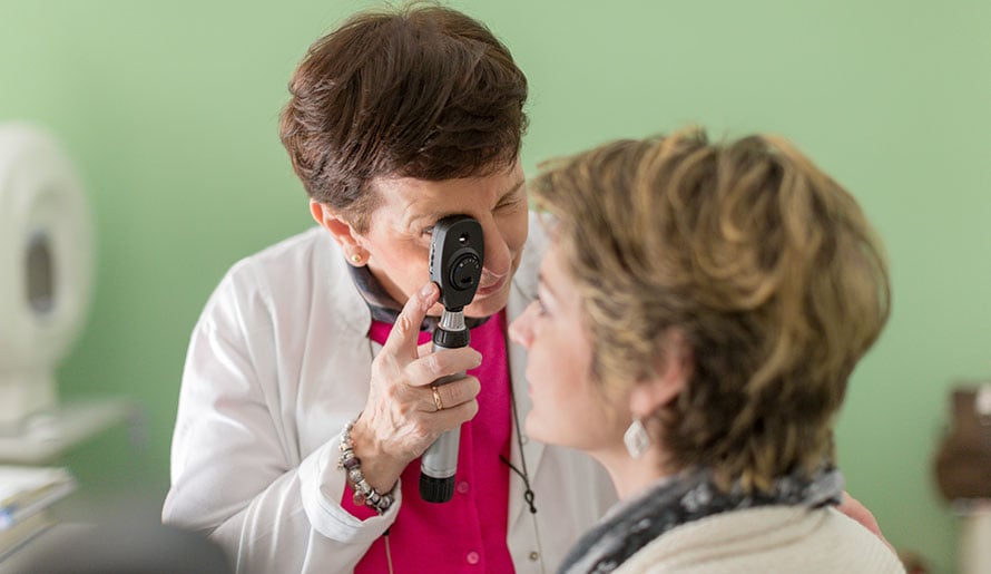
Ophthalmologists often detect cases of ocular melanoma during routine dilated eye exams. If a physician suspects that a patient might have this malignancy, they will likely order further testing, possibly including:
- Biopsy – A provider collects a small tissue sample and sends it to a pathologist for lab testing.
- Fluorescein angiography – A provider injects dye into the patient’s arm. Once the dye travels to the patient’s eye, the provider uses a special camera to take pictures of the inside of the eye.
- Fundus autofluorescence – A provider takes pictures of the patient’s eye using a special camera, which shows damaged areas as small points of light in the resulting image.
- Optical coherence tomography – A provider uses light waves to take cross-section photographs of the patient’s retina, allowing them to see each layer in greater detail.
- Ultrasound imaging – A provider administers eye drops to numb the patient’s eye, then gently places a small probe onto the eye’s surface. The probe sends and receives sound waves, and the echoes create an image of the eye’s interior.
If a physician determines that a patient does have ocular melanoma, they’ll likely order additional tests to determine whether the malignancy has metastasized (spread) to another area of the body. When ocular melanoma does metastasize, it most commonly spreads to the liver.
Ocular melanoma treatment
Ocular melanoma treatment will vary based on the patient’s age and overall health as well as the extent and location of their cancer. If a patient has a small eye melanoma, radiation therapy, laser therapy or photodynamic therapy may be recommended to shrink or destroy the cancer. An active surveillance approach may also be taken if the melanoma has not spread and is not causing symptoms. In more advanced cases, surgery may be recommended to remove part or all of the eyeball.
The Moffitt Cancer Center difference for ocular melanoma
If you’re concerned that you might have ocular melanoma, you can rely on the experienced team at Moffitt Cancer Center for diagnosis and treatment. Patients in our Cutaneous Oncology Program benefit from the expertise of surgical oncologists, plastic surgeons, radiation oncologists and other experts who specialize in ocular melanoma as well as other types of melanoma and skin cancer. We’re also proud to operate the Donald A. Adam Melanoma and Skin Cancer Center of Excellence, which is at the forefront of melanoma and skin cancer research.
Call 1-888-663-3488 or submit a new patient registration form online if you would like to speak with a Moffitt specialist. We welcome patients with or without referrals, and we’ll be sure to quickly connect you to a cancer expert.
References
American Academy of Ophthalmology: What Is Ocular Melanoma?
National Organization for Rare Disorders: Ocular Melanoma
