Esophageal Cancer
 Cancer that originates in the esophagus is fairly rare, accounting for about 1% of cancer cases in the U.S. As the name denotes, esophageal cancer affects the hollow, muscular tube that connects the mouth to the stomach and carries swallowed food and liquids through the throat and into the digestive tract. With this type of cancer, the tumor often develops in the cells that make up the moist lining of the esophagus (mucosa).
As esophagus cancer progresses, it may spread beyond the esophageal lining and invade the outer layers of the esophagus and nearby lymph nodes. After entering the lymphatic system, esophageal cancer can potentially travel throughout the body and spread to distant organs and tissues. This type of cancer typically grows slowly, and it may be many years before the symptoms become noticeable. However, once the symptoms begin, the cancer progresses rapidly.
What are the types of esophageal cancer?
Cancer of the esophagus is categorized based on the type of cells in which the malignancy originated. The two main types are:
- Squamous cell carcinoma – Forms in the thin, flat cells in the esophageal lining, usually in the upper two-thirds of the esophagus (near the throat)
- Adenocarcinoma – Forms in the mucus-producing glandular cells in the esophageal lining, usually in the lower third of the esophagus (near the stomach). While squamous cell carcinoma is the most prevalent type of esophageal cancer worldwide, adenocarcinoma is the most common type of esophageal cancer in the U.S.
What are the symptoms of esophageal cancer?
Oftentimes, the initial warning signs of esophageal cancer – if any – are mild, nonspecific and easily overlooked. In many cases, the first symptom is difficulty swallowing (dysphagia), either from coughing or choking that interferes with swallowing, or a sensation that food has become lodged in the throat. As esophageal cancer progresses, its symptoms typically worsen.
In addition to dysphagia, some other warning signs of esophageal cancer include:
- Pain or a burning sensation behind the breastbone in the middle of the chest
- Heartburn
- Indigestion
- Decreased appetite
- Unexplained weight loss
- Blood in the stool (caused by esophageal bleeding)
- A palpable lump under the skin in the chest
If esophageal cancer spreads beyond the esophagus, it can produce a variety of other symptoms depending on which organs or tissues are affected. Some additional signs of esophageal cancer include:
- Hiccups
- Chronic hoarseness or coughing
- Back pain
- Hypercalcemia and bone pain
- Respiratory fistulas
It’s important to note that, in addition to esophageal cancer, many of these symptoms can also be caused by other, less serious conditions. Therefore, in order to ensure prompt and effective treatment – which can lead to a better outcome and quality of life – it’s best to consult with a physician about any unusual health changes as soon as possible.
What causes esophageal cancer?

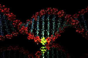
Esophagus cancer is relatively uncommon, and its precise causes are still unclear. However, scientists have identified a causal link between this form of cancer and damaged deoxyribonucleic acid (DNA) in esophageal mucosal cells.
DNA is a chemical that serves as a carrier of genetic information, which controls cellular function and provides instructions on when specific cells should replicate and die. Genes that instruct cells to grow, divide and remain alive are called oncogenes. Genes that instruct cells to stop dividing and die at the appropriate time are called tumor suppressor genes.
Damaged DNA can send faulty instructions that inadvertently “turn on” oncogenes or “turn off” tumor suppressor genes. This can cause some cells to divide very rapidly and others to live beyond their normal lifespan. As a result, an overabundance of cells may be created, and some of the excess cells may bind together and form tumors.
What are the risk factors for esophageal cancer?
Certain traits – both acquired and inherited – can affect the integrity of the cellular DNA in the esophageal mucosa and thereby increase the risk of esophageal cancer. A known risk factor for esophageal cancer is esophageal irritation, or chronic inflammation of the esophageal lining. This type of damage to the esophagus may be caused by gastroesophageal reflux disease (GERD), which is often a precursor to a condition known as Barrett’s esophagus. In the U.S., the most common risk factor for esophagus cancer is gastroesophageal reflux and its association with Barrett’s esophagus. However, Barrett’s esophagus can also develop without the presence of GERD.
Other conditions that increase a person’s risk for developing esophageal cancer include achalasia, a condition in which the muscles in the lower esophagus fail to relax, preventing food from passing into the stomach, and Plummer-Vinson syndrome, an uncommon condition that causes difficulty swallowing. Esophageal webs, which are thin layers of fibrous tissue that form in the throat, have also been associated with an increased risk for esophageal cancer, as has esophagus scarring from swallowing lye.
Additionally, some environmental factors and lifestyle choices may increase a person’s risk for developing esophagus cancer. Here are a few:
- Esophageal trauma – A direct blow or other injury to the esophagus
- Exposure to cancer-causing substances – Sustained contact with chemical carcinogens, such as acetaldehyde and polycyclic aromatic hydrocarbons, or environmental carcinogens, such as silica and asbestos
- Certain habits – Excessive consumption of tobacco products, alcoholic beverages, maté or very hot liquids
- Nutritional deficiencies – A vitamin or mineral imbalance or insufficient intake of fruits or vegetables
- Certain medications – Long-term use of nonsteroidal anti-inflammatory drugs (NSAIDs) or H2-receptor antagonists
- Obesity – Excess body weight or fatty tissue
- Genetics – Inherited DNA mutations (research suggests that esophageal cancer does not run in families, so the role of inherited DNA mutations in its development is believed to be minor)
To the extent possible, it is important to manage these risk factors because DNA damage can affect not only the cells in the esophageal lining but also other cells throughout the body. In fact, scientists believe cellular DNA damage contributes to the development of many types of cancer.
Risk of esophageal cancer by age
People between the ages of 45 and 70 have the highest risk of developing esophageal cancer, and fewer than 15% of cases are found in people younger than 55, according to the American Cancer Society. Moreover, men are three to four times as likely to have this type of cancer as women.
How is esophageal cancer diagnosed?

The diagnostic process for esophageal cancer usually begins with an imaging test, such as an upper gastrointestinal study with a barium swallow (esophagram). A harmless chalky liquid, barium can be swallowed to coat the inner walls of the esophagus before X-rays are taken to enhance the clarity of the resulting images. Other imaging tests, such as magnetic resonance imaging (MRI), computed tomography (CT) with contrast and positron emission tomography (PET) scans, may be used as well.
In images, early-stage esophageal cancer may resemble small round bumps or raised flat areas (plaques). More advanced esophageal cancer may appear as large irregular or narrowed areas. If images reveal possible esophageal cancer, the diagnostic process may continue with:
- An esophagoscopy – A thin, flexible tube with an attached light and tiny camera (scope) is passed down the patient’s throat and into his or her esophagus. High-resolution images captured by the camera are displayed on an external monitor in real time, allowing a physician to view the interior of the esophagus in detail.
- A biopsy – During an esophagoscopy or other endoscopic procedure, a physician passes a special instrument through the scope to collect a sample of esophageal tissue. The sample can then be examined under a microscope for evidence of cancer.
- Blood testing – A complete blood count (CBC) can detect anemia (a lower-than-normal red blood cell count), which may result from a bleeding esophageal tumor. A liver enzyme test can check the function of the liver, which may be affected by metastasized esophageal cancer.
If a diagnosis of esophageal cancer is confirmed, further tests may be performed to determine if and how far the cancer has spread. This information, which is known as the cancer’s stage, is essential for developing an appropriate treatment plan.
What are the stages of esophageal cancer?
In many cases, esophageal cancer is staged pathologically through an examination of tissue samples obtained during a surgical procedure. The staging process is complex and takes many factors into account. When determining a cancer’s stage, many physicians utilize the American Joint Committee on Cancer (AJCC) TNM system, which is based on three key pieces of information:
- Tumor size (T) – The extent to which cancerous cells have spread beyond the esophageal lining to the outer layers of the esophagus
- Lymph node involvement (N) – The extent to which cancerous cells have invaded nearby lymph nodes
- Metastasis (M) – The extent to which cancerous cells have metastasized to distant lymph nodes or organs
After evaluating the TNM information, a physician can classify the cancer into a stage. In simplified terms, the stages of esophageal cancer are:
- Stage 0 – Noncancerous but abnormal esophageal cells (high-grade dysplasia) have been detected, which could potentially progress into cancer.
- Stage 1 – Esophageal cancer cells have grown into the inner lining of the esophageal wall.
- Stage 2 – Esophageal cancer cells have spread into the main muscular layer of the esophagus.
- Stage 3 – Esophageal cancer cells have penetrated the outer wall of the esophagus and invaded nearby lymph nodes or organs.
- Stage 4 – Esophageal cancer cells have metastasized to distant lymph nodes or organs.
How is esophageal cancer treated?
When detected in its early stages, esophageal cancer is very treatable. In determining the most appropriate treatment approach, a key consideration is whether the cancer can be completely removed with surgery (resected), which is the most common form of esophageal cancer treatment. To make this determination, a physician will assess the location of the cancer and the extent of its spread, and also evaluate whether the patient is strong enough to undergo surgery.
All stage 0, 1 and 2 esophageal cancers are potentially able to be removed with surgery, as are most stage 3 esophageal cancers that have not spread to the trachea (windpipe), aorta (large blood vessel coming from the heart) or spine. Esophageal cancers that have spread to these vital structures or to distant lymph nodes or organs are usually not able to be removed with surgery. In these situations, other treatment approaches such as chemotherapy or radiation therapy may be discussed.
In general, the main treatment options for esophageal cancer are:
Surgery
The procedure most commonly performed to address esophageal cancer is an esophagectomy, which involves the removal of all or a portion of the esophagus, some nearby lymph nodes and a portion of the stomach. If the esophagus is completely removed, a passageway for food may be created from a section of the stomach or large intestine. If the esophagus is partially removed, the remaining part of the stomach may be repositioned in the chest or neck and connected to the remaining part of the esophagus. Until the patient’s swallowing function is restored, a feeding tube may be temporarily used during the recovery period to deliver nutrition directly to the stomach.
Radiation therapy
Radiation therapy involves the precise delivery of high-energy X-rays or particles directly to a tumor to destroy cancerous cells. This treatment may be administered prior to surgery to shrink a tumor and make it easier to remove, after surgery to target residual cancer cells or as a main form of treatment if surgery is not appropriate. For enhanced effectiveness, radiation therapy is sometimes combined with chemotherapy (a treatment approach known as chemoradiation).
The two main types of radiation therapy used to treat esophageal cancer are:
- External-beam radiation therapy (EBRT) – The most common form of radiation treatment for esophageal cancer, external radiotherapy involves the delivery of high-energy beams produced by an X-ray machine called a linear accelerator, which moves around the patient’s body without touching him or her. The beams, which can be delivered from any angle, are shaped to precisely match the contours of the tumor. The length of treatment depends on several factors, including the type and stage of the cancer. EBRT is often performed as an outpatient procedure over the course of two to 10 weeks, with one treatment session administered daily for five consecutive days, followed by a two-day break.
- Internal radiation therapy (brachytherapy) – After passing a scope down the patient’s throat and into his or her esophagus, a physician places tiny radioactive seeds very close to the cancer site. Because the energy must travel only a short distance to reach the targeted tumor, the effect on nearby healthy tissues is minimized. The radioactive source is removed a short time later. While brachytherapy is not well suited for treating a large area, it may be used to shrink a tumor, which can ease swallowing and relieve other symptoms of advanced esophageal cancer.
Chemotherapy
When used to address esophageal cancer, chemotherapy is usually administered through the bloodstream (systemic treatment) for widespread cancer cells that have spread or metastasized. Chemo involves the use of powerful medications, which are swallowed or injected into a vein. After entering the bloodstream, the medications can circulate throughout the body to reach cancerous cells that have spread to distant organs and tissues.
Chemotherapy can be very effective for destroying cancerous cells because the medications used specifically target rapidly dividing cells (rapid cell division is a hallmark of cancer). However, some healthy cells also share this characteristic. For instance, cells in the bone marrow, hair follicles and intestinal lining naturally divide very quickly. Because chemotherapy medications cannot differentiate between rapidly dividing cancer cells and rapidly dividing healthy cells, this treatment can potentially destroy some healthy cells. Depending on which cells are affected, a number of side effects can result, such as fatigue, easy bruising, increased risk of infection, anemia, hair loss, nausea and vomiting.
For this reason, chemotherapy is usually administered in cycles, with each treatment period followed by a break to allow the body some time to recover from any side effects. In general, chemotherapy cycles span two to four weeks, and many patients receive several cycles of treatment.
As research continues and more is being learned about the unique characteristics of cancer cells, new medications are continually being developed to precisely target these characteristics and fine-tune cancer treatment, thereby reducing the likelihood of side effects. Known as targeted therapy, this approach may be used along with or in place of traditional chemotherapy.
Can esophageal cancer be prevented?

As of yet, no one knows for sure what causes esophageal cancer to develop, and therefore how it can be prevented. Most likely, cancerous changes in the esophageal lining result from a complex interplay of a variety of physical, lifestyle and environmental factors. Some of these factors, such as smoking, are already well established – and can be controlled. This is significant because, even though there is no sure way to prevent esophageal cancer, there are ways to reduce the risk. Most involve healthy habits that can be used in daily life, such as:
- Avoiding tobacco use, which is a known risk factor not only for esophageal cancer but also for many other types of cancer
- Limiting alcoholic beverages, maté and very hot liquids
- Eating a nutritious, well-balanced diet that is rich in a variety of fruits and vegetables
- Exercising regularly
- Losing weight, if necessary, and maintaining a healthy body weight
- Learning about the risk factors for esophageal cancer and discussing them with a physician
Another important preventive measure is to seek prompt medical attention for persistent heartburn, which may be a sign of gastroesophageal reflux disease (GERD), a known risk factor for esophageal cancer. The role of GERD in the development of esophageal cancer is well understood. Normally, the lower esophageal sphincter (LES), a muscle at the end of the esophagus, opens to allow food to pass into the stomach, then closes to prevent a backflow of digestive acids. If the LES does not function properly, the resulting backwash (acid reflux) can irritate the lining of the esophagus. Studies have identified a link between chronic esophageal irritation and the development of adenocarcinoma of the esophagus.
Esophageal cancer treatment at Moffitt Cancer Center
Moffitt Cancer Center offers a full range of the latest treatment options for esophageal cancer, including esophagectomy and other surgical procedures performed with the robotic assistance of the da Vinci Surgical System, radiation therapy specifically for gastrointestinal cancers, chemoradiation and novel combinations of chemotherapy medications.
The multispecialty team in our renowned Gastrointestinal Oncology Program includes board-certified, fellowship-trained surgeons as well as many highly experienced gastroenterologists, medical oncologists, endoscopic oncologists, radiation oncologists, radiologists, pathologists, nurses, dietitians and other supportive care specialists. Working together, these medical professionals develop comprehensive and highly tailored treatment plans for our patients, with each team member contributing unique and valuable insight.
As a high-volume cancer center, Moffitt treats more uncommon cancers in a single day than many other cancer treatment centers see in a lifetime. Because our physicians treat many patients with esophageal cancer, we’ve acquired extensive expertise with regard to this relatively uncommon form of cancer. The depth of our esophageal cancer specialists’ experience directly translates to more accurate diagnoses and more effective treatment plans, as well as enhanced outcomes and quality of life for our patients.
In addition to unparalleled expertise, Moffitt’s approach to esophageal cancer treatment provides many other advantages for our patients, including:
The ultimate in multispecialty cancer treatment
Many cancer centers tout a multispecialty approach, but ours is one of the few in which all team members are available in one place. This not only brings a high level of collaboration; it also ensures a streamlined patient experience.
At Moffitt, we view our patients as key members of our team. As such, we ensure that they are fully informed, expertly guided and highly involved at every step of the way, from esophageal cancer prevention to survivorship. And our patients never have to worry about requesting referrals or traveling to different clinics – we provide everything they need in a single, convenient location. That’s one reason why thousands of patients – not only from Florida but also from across the nation and around the world – choose Moffitt for their cancer treatment.
The benefits of our groundbreaking research
The scientists, researchers and clinicians at Moffitt have contributed to some of the most significant breakthroughs in esophageal cancer treatment to date. By offering suggestions based on the results of our recent research, our team is able to recommend the latest and most effective strategies for fighting esophageal cancer.
Additionally, through our robust portfolio of clinical trials, our patients have access to promising new treatment options that are not yet available in other settings. Many innovative and novel therapies are available at Moffitt several years before they become the new standard of care, allowing our patients to be among the first to benefit. Our clinical trials incorporate compassionate patient care while evaluating the leading-edge developments in cancer treatment, sometimes offering options for even very aggressive or difficult-to-treat esophageal tumors.
As a National Cancer Institute-designated Comprehensive Cancer Center, Moffitt is nationally recognized for our extensive research efforts. We're passionate about advancing the treatment of all forms of cancer, including esophageal cancer, and we’re equally passionate about developing the clinicians of tomorrow. In fact, Moffitt trains more students in the field of oncology than all other institutions in the state of Florida combined. We remain steadfastly committed to continuing these efforts at our current pace – right up until the day we find a cure.
The intangible human touch
At Moffitt, we firmly believe that basic human kindness can have a profound positive effect in a health care setting, and we see evidence of this every single day. We’re committed to improving esophageal cancer treatment not only through science and technology but also through empathy and compassion. When blended together, these four essential components – science, technology, empathy and compassion – form a powerful combination that creates an optimal environment for healing – and ultimately saves lives.
At Moffitt, planning for your personalized treatment begins even before you step in the door. When you reach out to us, we’ll provide you with rapid access to a cancer expert so that we can start treatment as soon as possible. To request a consultation with an esophageal cancer expert in the Gastrointestinal Oncology Program at Moffitt Cancer Center, call 1-888-663-3488 or complete a new patient registration form.
References
National Cancer Institute: Lymphatic System
Cancer.Net: Esophageal Cancer Risk Factors
American Cancer Society: Esophageal Cancer Risk Factors
National Cancer Institute: Esophageal Cancer
Support the Future of Gastrointestinal Oncology Research and Treatment
When you support Moffitt Cancer Center, you help make breakthrough gastrointestinal research and innovative treatments possible.
Give now to support the Gastrointestinal Oncology Program. For more information, call toll-free 1-800-456-3434, ext. 1403.
