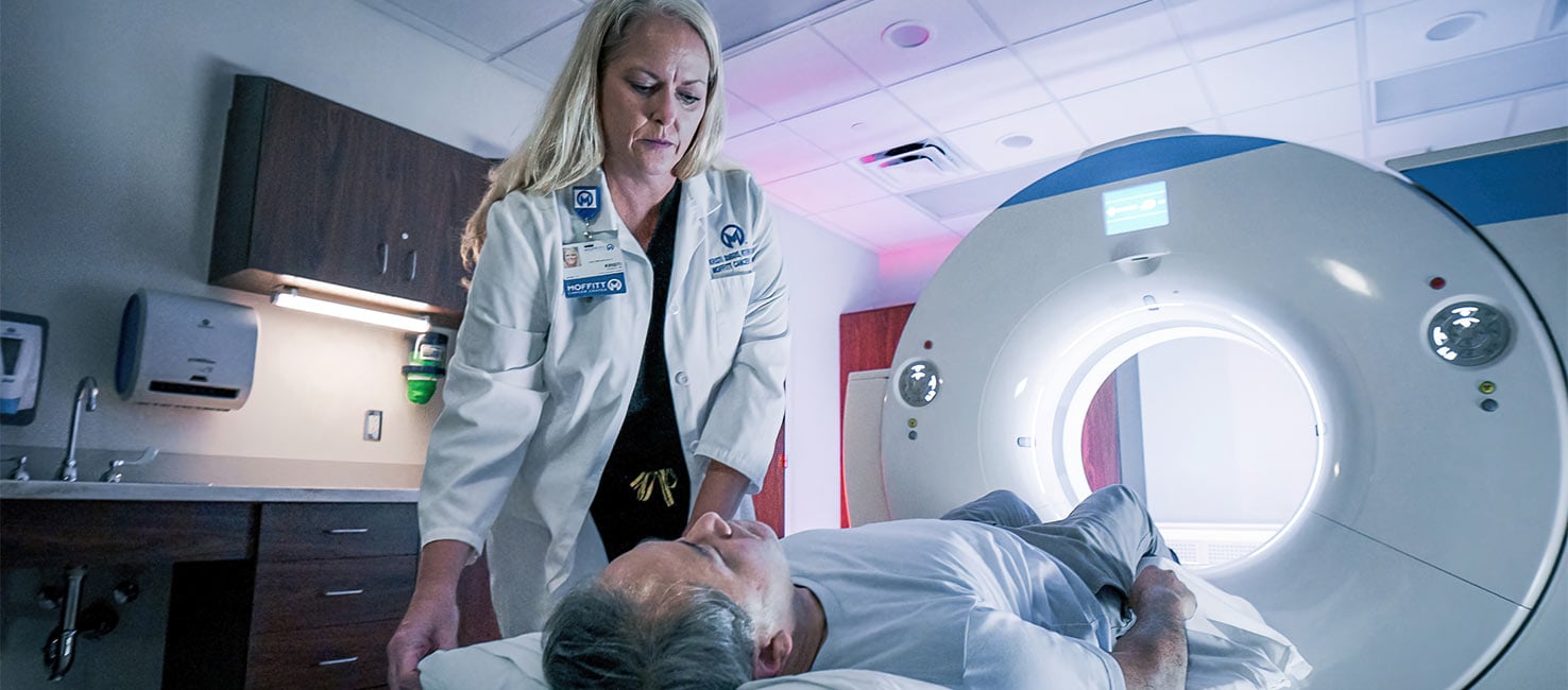
Advanced Imaging for Local Evaluation of Urinary Bladder Cancer: The Role of MRI
More than 80% of newly diagnosed urinary bladder cancer are non-muscle invasive (NMIBC). This cancer type has a high recurrence rate. Many patients with non-muscle-invasive bladder cancer will experience a relapse within the first five years of diagnosis.
It’s estimated about 30% of NMIBC patients are under-staged after TURBT and this increases to 45% if resection does not include detrusor muscle. Furthermore, about 1/3 of tumors that were clinically confined to the bladder were shown on final pathology following a radical cystectomy which demonstrates an extra-vesical extension.
The Vesical Imaging-Reporting and Data System (VI-RADS) scoring system was created to standardize the protocols for MR imaging of bladder cancer. The VI-RADS provides a structured reporting system and helps to improve communication between referring physicians and radiologists. It also provides a risk score for muscle layer invasion in bladder cancer.
The VI-RADS scoring system is based on a 5-point assessment scale:
- VIRADS 1: Muscle invasion is highly unlikely.
- VIRADS 2: Muscle invasion is unlikely to be present.
- VIRADS 3: The presence of muscle invasion is equivocal.
- VIRADS 4: Muscle invasion is likely
- VIRADS 5: Invasion of muscle and beyond the bladder is very likely.
The VI-RADS score system has been tested in multiple studies and has been shown to have high reproducibility and diagnostic accuracy for determining muscle layer invasion. It also showed high diagnostic validity and reliability in predicting urinary bladder muscle invasion, especially VI-RADS 4 and 5, and in excluding the muscle invasiveness in VIRADS 1. However, there is a percentage of bladder cancer with muscle invasion in VIRADS 2 (~25%) and VIRADS 3 (~50%) and this would require further modifications to enhance the diagnostic validity for these scores.
To prepare a patient for the MRI procedure, the urinary bladder should be performed before TURBT.
- Eight weeks after TURBT, follow up with a bladder biopsy, or intravesical treatment to avoid reactive changes in the urinary bladder wall.
- A cystoscopy or removal of Foley catheter a week later because the air in UB causes artifact on DWI/ADC.
- Adequate bladder distension is required about one to two hours before imaging. The patient should start drinking 500–1000 ml of water 30 min before the examination.
Application of VIRADS in clinical practice
Assessment of detrusor muscle invasiveness and differentiation of NMIBC and MIBC.
For patients with NMIBC:
- Plan surgically feasible therapeutic TURBT.
- Improve the selection of patients who are candidates for Re-TURBT.
- In the surveillance setting, for patients with NMIBC who require frequent cystoscopy to detect possible recurrence, MRI could be used as a noninvasive follow-up alternative.
For patients with MIBC:
- Stage tumors that benefit from neoadjuvant therapy.
- Use of MRI during chemotherapy: earlier prediction of treatment failure
In summary, the role of MRI in urinary bladder cancer management is evolving and recent data show it is an important impact on patient management.
If you'd like to refer a patient to Moffitt Cancer Center, complete our online form or contact a physician liaison for assistance. As part of our efforts to shorten referral times as much as possible, online referrals are typically responded to within 24 - 48 hours.

