
Astrocytoma Treatment
Astrocytoma is a type of glioma that develops from or resembles astrocytes, star-shaped cells that support nerve cells in the brain and spinal cord. Treatment is individualized based on the tumor’s location, grade and molecular characteristics, with goals that may include removing as much tumor as safely possible, slowing or controlling further growth, preserving neurological function and managing symptoms.
At Moffitt Cancer Center, patients with astrocytoma receive specialized care through the Neuro-Oncology Program. Our neurosurgeons, neuro-oncologists, radiation oncologists, neuropathologists, neuroradiologists and other specialists collaborate to evaluate each tumor and develop an individualized treatment plan.
Quick facts: treating astrocytoma
- Surgery is often the foundation of treatment. When an astrocytoma can be safely approached, the goal is typically maximal safe resection—removing as much tumor as possible without unnecessarily compromising important neurological functions.
- Additional treatment depends on the tumor. Radiation therapy and chemotherapy may be recommended after surgery for certain astrocytomas, while carefully selected patients with lower-grade tumors may initially undergo active surveillance or receive targeted therapy instead.
- Molecular testing plays a central role. Modern astrocytoma classification incorporates molecular features, particularly isocitrate dehydrogenase (IDH) status. These findings help establish the precise diagnosis and grade and can directly affect treatment decisions.
- Treatment options continue to advance. Surgery, increasingly precise radiation delivery techniques, chemotherapy and molecularly targeted therapies provide several ways to control astrocytoma. For example, the IDH inhibitor vorasidenib is an FDA-approved option for certain grade 2 IDH-mutant astrocytomas following surgery.
Surgery for astrocytoma
Surgery is an important part of treatment for many astrocytomas. To plan the procedure, a neurosurgeon who specializes in brain tumors evaluates the tumor’s size and location as well as its relationship to areas responsible for speech, movement, sensation and other essential functions. Moffitt offers a range of neurosurgical treatment approaches, including minimally invasive cranial surgery and complex awake craniotomy.
When is surgery a treatment option for astrocytoma?
Surgical treatment may be recommended when an astrocytoma can be accessed without creating an unacceptable risk of neurological impairment. Rather than simply removing as much tissue as possible, the goal is maximal safe resection—removing the greatest amount of tumor that can be safely taken out while preserving healthy brain tissue and neurological function.
Surgery can also provide tissue for neuropathological and molecular analysis, which is essential for establishing an accurate diagnosis and guiding further treatment. In some cases, the tumor’s location makes extensive removal unsafe. A biopsy or partial resection may then be appropriate, followed by other treatment or careful monitoring depending on the tumor’s characteristics.

ACS Surgical Quality Partner for 30+ Years
Continuously Accredited by the American College of Surgeons Commission on Cancer since 1989 for our commitment to providing comprehensive, high-quality and multispecialty patient-centered care.
What are the different types of astrocytoma surgery?
The surgical approach is tailored to the tumor’s location, growth pattern and relationship to critical brain structures. Options may include:
Craniotomy
A craniotomy involves temporarily removing a section of the skull, known as a bone flap, so the neurosurgeon can access the tumor. The surgeon removes as much tumor as safely possible and then replaces the bone flap. Advanced imaging, navigation systems and other technologies can help distinguish tumor tissue from healthy brain tissue and guide the resection.
Awake craniotomy and intraoperative brain mapping
An awake craniotomy may be considered when an astrocytoma is close to an area of the brain involved in speech, movement, sensation or another critical function. During part of the operation, the patient is awake and able to respond to questions or perform specific tasks while the surgical team stimulates and maps areas of the brain.
This real-time feedback helps the neurosurgeon identify functional tissue that should be preserved while determining how much tumor can safely be removed. Moffitt performs awake craniotomies as part of our specialized brain tumor surgery program.

Stereotactic biopsy
If removing the tumor is not initially appropriate, a neurosurgeon may perform a stereotactic biopsy. Using imaging and computer-guided navigation, the surgeon obtains a small sample of tumor tissue for examination. Neuropathological and molecular testing of this tissue can establish the diagnosis and provide information needed to select treatment.
What are the risks and possible side effects of astrocytoma surgery?
The potential effects of brain surgery depend heavily on where the tumor is located and which surrounding structures are involved. The surgical team weighs these risks against the potential benefits before recommending a procedure. Possible side effects of astrocytoma surgery include:
- Headache and discomfort – Pain and swelling can occur after a craniotomy. Medications and careful postoperative monitoring can help keep these symptoms under control while healing progresses.
- Temporary neurological changes – Weakness, numbness, difficulty speaking, problems with coordination or other changes can occur if treatment affects nearby brain tissue. Neurological assessments after surgery help identify changes promptly, and physical, occupational or speech therapy may be incorporated into recovery.
- Seizures – Brain tumors and brain surgery can increase seizure risk. The healthcare team monitors for seizure activity and may prescribe antiseizure medication when clinically appropriate.
- Bleeding, swelling or infection – These are recognized risks of cranial surgery. The surgical team monitors the patient closely during the early recovery period so complications can be identified and treated promptly.
- Fatigue and reduced stamina – Significant tiredness is common during early recovery. Gradually increasing activity according to the surgical team’s recommendations can help the patient regain strength without overexertion.
What is the recovery process like after astrocytoma surgery?
Recovery varies based on the extent and location of the surgery, the patient’s neurological function before the procedure and whether additional treatment is planned. A patient may spend several days in the hospital while the healthcare team monitors neurological function, manages pain and evaluates the surgical site.
The recovery process then continues at home over the following weeks. Some patients gradually return to their usual activities with relatively little assistance, while others may benefit from physical therapy, occupational therapy, speech-language therapy or neurocognitive rehabilitation. Follow-up imaging is generally performed to assess the extent of tumor removal and establish a baseline for future monitoring.
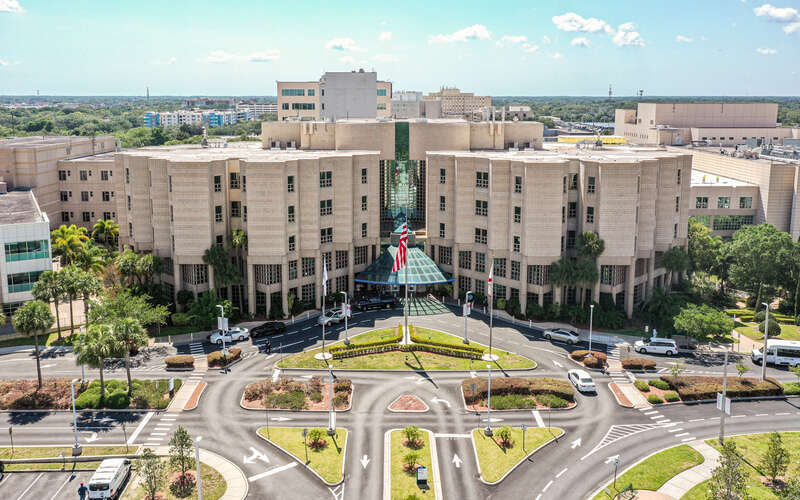
#1 Cancer Hospital in Tampa
Moffitt is the top-ranked cancer hospital in Tampa and central Florida and ranked No. 16 in the nation in Newsweek’s “America’s Best Cancer Hospitals."
Chemotherapy for astrocytoma
Chemotherapy is a systemic treatment that uses medications to damage cancer cells or interfere with their ability to grow and divide. Unlike surgery and radiation therapy, which target a specific area, systemic medications circulate through the body.
Treating a brain tumor with systemic medication presents a particular challenge because of the blood-brain barrier, a network of tightly connected blood vessels that protects the brain by limiting the passage of many substances from the bloodstream into brain tissue. To be effective against an astrocytoma, a systemic drug must be able to reach the tumor in a therapeutically useful amount. Temozolomide, an oral chemotherapy medication frequently used to treat gliomas, penetrates the central nervous system.
When is chemotherapy used for astrocytoma?
Chemotherapy for astrocytoma is generally considered after the tumor has been surgically removed or biopsied and its molecular features and grade are known. The need for chemotherapy varies considerably.
For some grade 2 IDH-mutant astrocytomas, careful surveillance or an IDH-targeted medication may allow radiation therapy and chemotherapy to be deferred. When additional treatment is indicated for grade 2 disease, radiation therapy may be combined with chemotherapy. Grade 3 IDH-mutant astrocytomas are generally treated with radiation therapy followed by temozolomide, while treatment of grade 4 IDH-mutant astrocytoma is individualized according to the tumor’s characteristics and the patient’s circumstances.
Chemotherapy may also be considered if an astrocytoma progresses or returns following earlier treatment.
Which chemotherapy drugs are used for astrocytoma?
The chemotherapy regimen for astrocytoma depends on the tumor’s grade, molecular characteristics and previous treatment. Options can include:
- Temozolomide (Temodar) – This oral chemotherapy medication damages DNA within tumor cells, interfering with their ability to reproduce. Temozolomide penetrates the central nervous system and is an important component of treatment for many higher-grade diffuse gliomas.
- PCV chemotherapy – This combination of procarbazine, lomustine and vincristine may be considered in certain treatment plans for grade 2 IDH-mutant astrocytoma. The three medications interfere with tumor growth through complementary mechanisms, although the regimen can produce more substantial side effects than temozolomide and is not appropriate for every patient.
The healthcare team considers the potential benefits of each regimen along with the patient’s previous treatments, overall health and ability to tolerate therapy.
 The Only Florida-based NCI-designated Comprehensive Cancer Center
Recognized for its scientific excellence, Moffitt is the only National Cancer Institute-designated Comprehensive Cancer Center based in Florida.
What are the risks and possible side effects of chemotherapy for astrocytoma?
Side effects vary according to the medication, dose and individual response. The healthcare team monitors blood counts and other health indicators throughout treatment so supportive care or dose adjustments can be provided when needed. Possible side effects of chemotherapy for astrocytoma include:
- Fatigue – Chemotherapy and changes in blood cell production can contribute to low energy. Planned rest, appropriate physical activity and evaluation for treatable causes such as anemia can help manage fatigue.
- Nausea or reduced appetite – Temozolomide and other chemotherapy medications can cause digestive symptoms. Antinausea medications are often used, and nutrition specialists can provide strategies for maintaining adequate food and fluid intake.
- Low blood cell counts – Chemotherapy can temporarily reduce white blood cells, red blood cells or platelets. Regular blood tests allow the healthcare team to monitor these changes and adjust treatment or provide supportive medications when necessary.
- Increased infection risk – A reduction in infection-fighting white blood cells can make some patients more susceptible to illness. The treatment team provides instructions about infection precautions and when a fever or other symptom warrants prompt evaluation.
- Constipation or other digestive changes – Certain medications, including some components of PCV, can affect gastrointestinal function. Dietary changes, hydration and medications may be recommended based on the specific symptom.
How does chemotherapy for astrocytoma affect daily life, and what is the recovery process like?
Temozolomide is taken by mouth according to a prescribed schedule. Depending on the treatment plan, it may be given during a course of radiation therapy or in repeating cycles with treatment days followed by recovery periods. PCV incorporates oral medications as well as vincristine, which is administered intravenously.
Many patients can continue at least some usual activities during treatment, although fatigue and other side effects may fluctuate throughout each cycle. Blood tests and follow-up visits are used to assess treatment tolerance, and the healthcare team can adjust doses, delay a cycle or provide supportive treatment when needed.
Radiation therapy for astrocytoma
Radiation therapy uses precisely directed high-energy beams to damage the DNA of tumor cells, preventing them from continuing to grow and divide. For astrocytoma, treatment is carefully planned to deliver an effective dose to the tumor and surrounding area at risk while limiting radiation exposure to healthy brain tissue.
Moffitt’s radiation oncologists work with medical physicists, dosimetrists and other specialists to develop individualized radiation plans. Moffitt offers several advanced radiation delivery technologies that may be considered for brain tumors.
How does radiation therapy treat astrocytoma?
Radiation therapy is frequently used after astrocytoma surgery when the tumor’s grade, molecular features or extent indicate a meaningful risk of continued growth. It can target microscopic tumor cells that cannot be seen or safely removed during surgery.
Radiation treatment may also be used when an astrocytoma cannot be completely removed due to its location or when surgery is not appropriate. The timing, radiation dose and treatment field are individualized according to the tumor’s characteristics and the patient’s previous treatments.
What radiation treatment techniques are available for astrocytoma?
External beam radiation therapy (EBRT), which delivers radiation to a tumor from a machine outside the body, is the primary form of radiation therapy used for diffuse astrocytomas. Different technologies and delivery techniques can be used to precisely target the treatment area, with the most appropriate approach depending on factors such as the tumor’s location, size and proximity to critical structures. Options may include:
Intensity-modulated radiation therapy (IMRT)
An advanced form of EBRT, IMRT can vary the intensity and shape of individual radiation beams, allowing the radiation dose to conform closely to complex treatment areas in the brain. This precision can help limit unnecessary radiation exposure to nearby healthy brain tissue and sensitive structures while delivering the prescribed dose to the treatment area.
Proton therapy
Proton therapy is a form of EBRT that uses protons rather than the X-rays, or photons, used in conventional radiation therapy. Unlike X-rays, proton beams can deposit much of their energy at the intended depth within the body and then stop, potentially reducing radiation exposure beyond the treatment target.
Moffitt offers proton therapy for brain tumors at the Richard M. Schulze Family Foundation Proton Therapy Center at Speros. Proton therapy is not necessary or preferable for every astrocytoma, so candidacy is determined through consultation with a radiation oncologist.
Stereotactic radiation delivery techniques
Stereotactic radiation techniques use highly focused external radiation beams to deliver radiation with substantial precision. These techniques are generally more applicable to small, well-defined treatment targets than to the broad infiltrative margins characteristic of many diffuse astrocytomas. In selected circumstances, however, stereotactic radiation may have a role in treating a focal area of recurrent disease.
What are the possible side effects of radiation therapy for astrocytoma?
Side effects depend on the dose, treatment area and amount of healthy brain tissue exposed to radiation. Whenever possible, the radiation oncology team designs treatment to limit these effects, which may include:
- Fatigue – Tiredness can gradually increase during a multiweek course of treatment. Balancing rest with appropriate activity and maintaining adequate nutrition can help the patient manage changes in energy.
- Hair loss in the treatment area – Radiation therapy can affect hair follicles within the path of the beams. Hair may regrow after treatment, although the degree and timing vary with the radiation dose.
- Scalp irritation – Skin in the treatment area may become dry, tender or irritated. The radiation team can recommend appropriate skin care products and advise the patient about products or practices to avoid.
- Headache, nausea or brain swelling – Radiation therapy can temporarily cause inflammation within the treatment area. Medications may be used to manage nausea, headache or swelling when needed.
- Cognitive changes – Depending on the location and amount of brain tissue treated, radiation therapy can sometimes affect memory, concentration or processing speed. Careful treatment planning helps limit exposure to healthy tissue, and neurocognitive evaluation or rehabilitation may be useful if changes develop.
- Delayed tissue changes – Some side effects of radiation therapy can develop months or even years after treatment. Long-term follow-up and MRI surveillance help the healthcare team evaluate new symptoms or imaging changes and distinguish treatment effects from tumor progression.
When is the schedule for radiation therapy for astrocytoma, and what is the recovery process like?
When conventionally fractionated radiation therapy is used, treatment is typically delivered five days per week over several weeks. The exact number of sessions and radiation dose depend on the astrocytoma’s grade, location and treatment goals.
Each treatment session is relatively brief, although additional time is needed for careful positioning. Side effects such as fatigue can build gradually and may persist for a period after the final treatment before improving. Follow-up visits and imaging allow the healthcare team to monitor recovery and evaluate the tumor’s response.
Targeted therapy and immunotherapy for astrocytoma
Molecular testing has fundamentally changed how diffuse gliomas are classified and treated. Rather than relying on how tumor cells look under a microscope alone, neuropathologists analyze specific genetic and molecular features that can help establish the diagnosis, determine the tumor’s grade and identify potential treatment targets.
Which biomarkers are tested before targeted therapy is considered for astrocytoma?
The specific molecular tests performed can vary, but several findings are particularly important in the evaluation of a suspected diffuse astrocytoma:
- IDH1 and IDH2 mutations – IDH status is central to modern classification of adult-type diffuse gliomas. Adult-type astrocytomas with an IDH mutation are classified as astrocytoma, IDH-mutant, and may be assigned CNS WHO grade 2, 3 or 4.
- 1p/19q status – A tumor with an IDH mutation and combined deletion of chromosome arms 1p and 19q is classified as an oligodendroglioma rather than an astrocytoma. Testing therefore helps distinguish between these two major types of diffuse glioma.
- CDKN2A/CDKN2B – Homozygous deletion of these genes is an important molecular feature because its presence can establish a grade 4 designation in an IDH-mutant astrocytoma even without certain high-grade microscopic features.
- MGMT promoter methylation – MGMT status can provide information about how sensitive certain gliomas may be to alkylating chemotherapy such as temozolomide. Its clinical role varies according to the precise tumor type and treatment setting.
- Additional molecular alterations – Broader tumor profiling may identify less common changes that help refine the diagnosis, provide prognostic information or reveal a potential targeted treatment or clinical trial.
What targeted therapies are available for astrocytoma?
Vorasidenib (Voranigo) is an oral targeted medication that inhibits mutant IDH1 and IDH2 enzymes. The FDA has approved vorasidenib for patients age 12 and older with grade 2 astrocytoma or oligodendroglioma that has a susceptible IDH1 or IDH2 mutation following surgery, including biopsy, subtotal resection or gross total resection.
For selected patients with grade 2 IDH-mutant astrocytoma, vorasidenib may provide a treatment option when radiation therapy and chemotherapy can be deferred.
Like other systemic medications used for brain tumors, a targeted therapy must be capable of reaching its intended target within the central nervous system. This is one reason drug development for astrocytoma is particularly complex: the blood-brain barrier can prevent many medications circulating in the bloodstream from reaching brain tissue in adequate concentrations.
How is immunotherapy used for astrocytoma?
Immunotherapy is designed to help the immune system recognize or attack cancer cells. Although immune checkpoint inhibitors and other immunotherapies have become important treatments for several cancers, they are not currently routine standard treatment for IDH-mutant astrocytoma.
Researchers continue to study immune-based treatment strategies for brain tumors. For astrocytoma, these approaches remain largely investigational and are primarily available through clinical trials. Moffitt’s brain tumor research program includes studies of immunotherapies, vaccine therapies and other novel approaches.
Astrocytoma clinical trials at Moffitt Cancer Center
As a National Cancer Institute (NCI)-designated Comprehensive Cancer Center, Moffitt conducts clinical research aimed at improving the diagnosis and treatment of astrocytoma. Our brain tumor clinical trials investigate areas such as targeted therapies, new chemotherapy combinations and delivery methods, immunotherapies, vaccine therapies, radiation treatment approaches and surgical techniques. Patients who are eligible for a clinical trial may be able to receive an investigational therapy or treatment strategy before it becomes widely available.
Why should a patient consider a clinical trial for astrocytoma?
Despite significant advances in molecular classification and treatment, diffuse astrocytomas can be challenging to control because tumor cells may infiltrate surrounding brain tissue and because the blood-brain barrier limits the effectiveness of many systemic medications.
Clinical trials provide a structured way to evaluate strategies designed to address these challenges. Depending on the study and the patient’s eligibility, a trial may involve a molecularly targeted medication, immune-based treatment, new drug-delivery strategy, radiation approach or combination of therapies. Participation is voluntary, and the research team explains the potential benefits, risks and alternatives before enrollment.
Frequently asked questions (FAQs) about astrocytoma treatment
Astrocytoma treatment can differ substantially from one patient to another. The following answers address several common questions about how treatment decisions are made.
Can an astrocytoma be completely removed with surgery?
Sometimes. A neurosurgeon may be able to remove all of the tumor that is visible on imaging, particularly when the tumor has favorable surgical boundaries and is not intertwined with critical functional areas. However, diffuse astrocytoma cells can extend microscopically into surrounding brain tissue, so even a gross total resection does not necessarily mean that every tumor cell has been eliminated. Ongoing MRI surveillance remains important.
Is radiation therapy required after astrocytoma surgery?
Not always. The need for radiation therapy depends on factors such as the tumor’s grade and molecular characteristics, the extent of the resection, the patient’s age and any evidence of residual or progressive disease. For some carefully selected patients with grade 2 IDH-mutant astrocytoma, radiation therapy and chemotherapy may initially be deferred, with surveillance or targeted treatment with vorasidenib considered instead.
How does the grade of an astrocytoma affect treatment?
Grade helps describe the tumor’s biological behavior and is an important factor in determining how intensive treatment should be. Under current World Health Organization (WHO) classification, adult-type astrocytoma, IDH-mutant can be grade 2, 3 or 4. Grade 2 tumors may sometimes be managed with surgery followed by surveillance or targeted therapy, while grade 3 and 4 tumors generally require additional treatment such as radiation therapy and chemotherapy.
Is glioblastoma the same as a grade 4 astrocytoma?
Not under the current classification system. Astrocytoma, IDH-mutant, grade 4 and glioblastoma, IDH-wildtype, grade 4 are now classified as separate adult-type diffuse gliomas. This distinction is based on molecular features and is clinically important because the precise diagnosis can influence treatment planning and prognosis.
Why can’t every astrocytoma be completely removed?
Astrocytomas can grow into brain tissue responsible for essential functions such as speech, movement, vision or sensation. Diffuse tumor cells may also extend beyond the abnormality visible on an MRI. In these situations, attempting to remove every tumor cell could cause unacceptable neurological damage. Advanced approaches such as intraoperative brain mapping and awake craniotomy can help the neurosurgeon balance tumor removal with preservation of function.
Can an astrocytoma come back after treatment?
Yes. Even after extensive surgical removal, microscopic tumor cells can remain in surrounding brain tissue. The risk of recurrence varies according to the astrocytoma’s grade, molecular characteristics and response to treatment. Regular MRI surveillance helps the healthcare team identify changes that may require further evaluation or treatment.
Why choose Moffitt Cancer Center for astrocytoma treatment
Astrocytoma care requires specialized expertise in both brain tumor treatment and the complex molecular features that influence diagnosis and treatment planning. Moffitt’s Neuro-Oncology Program brings together neurosurgeons, neuro-oncologists, radiation oncologists, neuropathologists, neuroradiologists, neuropsychologists and other specialists who focus on brain and spinal tumors. Cases are reviewed collaboratively to help ensure that all appropriate surgical, systemic, radiation therapy and clinical trial options are considered.
As a high-volume cancer center, Moffitt also offers advanced neurological approaches ranging from minimally invasive cranial surgery to complex awake craniotomy, as well as precision radiation delivery technologies and an extensive brain tumor clinical trials program.
A patient who has already received an astrocytoma diagnosis elsewhere can also come to Moffitt for a second opinion. Specialized neuropathologists can review tumor tissue and molecular findings, while neuroradiologists and other members of the Neuro-Oncology Program can reassess imaging and treatment options. This comprehensive review can help confirm the precise diagnosis and grade and determine whether surgery, radiation therapy, chemotherapy, targeted therapy, active surveillance or a clinical trial should be considered.
If you would like to learn more about astrocytoma treatment, you can request an appointment with a specialist in the Neuro-Oncology Program at Moffitt by calling 1-888-663-3488 or submitting a new patient registration form online. We do not require referrals.
Treatment