
Ocular Melanoma Overview
Ocular melanoma is a rare cancer that develops in the pigment-producing cells (melanocytes) of the eye, which give the eye its color. Although melanoma most often forms in the skin, it can also develop in other areas of the body that have melanocytes, including the eye.
Most ocular melanomas arise in the middle layer of the eye (uvea), which comprises the iris, ciliary body and choroid. The iris is the colored part of the eye. The ciliary body is the circular structure that contains the ciliary muscle, which helps the eye focus by changing the shape of the lens. The ciliary body also produces aqueous humor, a clear fluid that fills the front part of the eye to maintain the eye’s internal pressure and provide nutrients to the cornea and lens. The choroid contains blood vessels that supply the retina. The majority of ocular melanoma cases occur in the choroid.
Early-stage ocular melanoma may progress without causing noticeable symptoms. In many cases, the condition is discovered during a routine eye examination. As the tumor grows, it may begin to affect vision and can potentially spread to other parts of the body, most commonly the liver. Early detection and specialized treatment are essential for preserving vision and improving the outcome.
According to the National Organization for Rare Disorders, ocular melanoma affects about five people per million each year in the United States, with roughly 2,500 new cases diagnosed annually. Even so, it is the most common type of primary eye cancer in adults.
What causes ocular melanoma?
Ocular melanoma develops when melanocytes within the eye undergo abnormal changes that cause them to grow and divide uncontrollably, eventually forming a tumor. The precise triggers of these DNA mutations are unknown. In many cases, ocular melanoma develops without a clearly identifiable cause.
Unlike skin melanoma, ocular melanoma is not strongly associated with ultraviolet (UV) light exposure. Instead, scientists believe that a combination of genetic susceptibility and environmental influences—which are not yet fully understood—may contribute to its development.
-

5 people per million
are diagnosed with ocular melanoma each year in the U.S.
-

About 2,500
new cases of ocular melanoma are diagnosed annually in the U.S.
What are the risk factors for ocular melanoma?
Several factors may increase the likelihood of developing ocular melanoma. These include:
- Advanced age (most cases are diagnosed after age 50)
- Certain genetic conditions, such as BAP1 tumor predisposition syndrome
- Fair skin or a tendency to sunburn easily
- Light eye color, such as blue, gray or green
- Ocular or oculodermal melanocytosis, a condition that causes increased pigmentation in and around the eye
- Preexisting eye moles (choroidal nevi)
Individuals with one or more of these risk factors may benefit from periodic eye exams to monitor changes in their eye health. It is also notable that many people diagnosed with ocular melanoma have no known risk factors.
Is ocular melanoma preventable?
There is no proven way to completely prevent ocular melanoma. Because the exact causes are unknown, prevention strategies are limited. That said, several measures can help support overall eye health and allow for early detection. These include:
- Having routine eye exams
- Monitoring known eye moles for changes in size or appearance
- Seeking a prompt medical evaluation for unusual eye symptoms or vision changes
- Wearing sunglasses that block UV light
Because ocular melanoma can develop without obvious symptoms, having regular eye examinations is especially important.

#1 Cancer Hospital in Tampa
Schedule an AppointmentWhat are the signs and symptoms of ocular melanoma?
Ocular melanoma may not cause symptoms in its earliest stages. When symptoms do occur, they often affect vision or the appearance of the eye. The signs can vary depending on the size and location of the tumor.
What are the early warning signs of ocular melanoma?
Often subtle, the early warning signs of ocular melanoma may include:
- Blurred vision
- Flashes of light in the visual field
- Partial loss of peripheral vision
- A small dark spot on the iris that increases in size
- Unusual changes detected during a routine eye exam
Because these symptoms can also occur with other eye conditions, a professional medical evaluation is important to ensure an accurate diagnosis.
What are common symptoms of ocular melanoma?
As the tumor grows, symptoms may become more apparent, such as:
- Changes in pupil shape
- Distorted vision
- Eye discomfort or pressure
- Floaters or spots in the field of vision
- Loss of part of the visual field
It is important to note that some patients may not experience symptoms, even if the tumor is relatively large.
What are the signs of advanced ocular melanoma?
Advanced ocular melanoma may produce more significant eye or vision changes. Signs may include:
- Bulging of the eye
- Increasing eye pain or pressure
- Severe vision loss in one eye
- A visible mass or swelling in the eye
These symptoms may indicate the tumor is growing or affecting the surrounding structures within the eye.
What are the signs that ocular melanoma has spread?
Ocular melanoma can potentially spread, or metastasize, from the eye to other parts of the body. The liver is the most common site of metastasis, although the lungs, bones and other organs can also be affected.
Signs that ocular melanoma has spread may include:
- Abdominal discomfort or swelling
- Bone pain
- Decreased appetite
- Fatigue
- Shortness of breath
- Unexplained weight loss
How is ocular melanoma diagnosed?
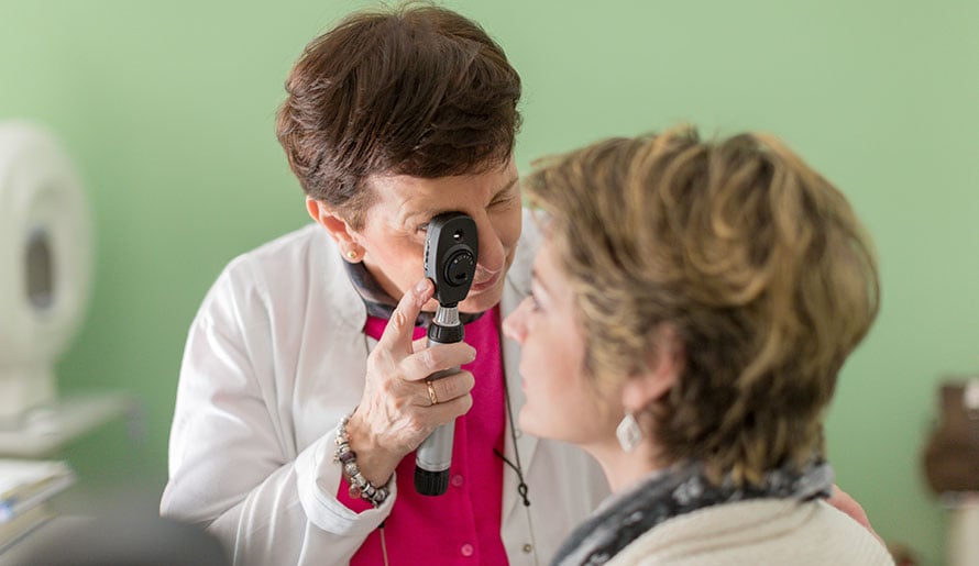
Ocular melanoma is typically diagnosed through a comprehensive eye exam and specialized imaging studies. During the evaluation, the physician will review the patient’s symptoms and medical history and perform a thorough examination of the eye.
What imaging tests are used to diagnose ocular melanoma?
Imaging plays a key role in diagnosing ocular melanoma and evaluating the location and size of the tumor. Common imaging tests include:
- Fluorescein angiography to visualize blood flow in the retina and choroid
- Fundus photography to document changes in the eye
- Ocular ultrasound, which uses sound waves to create detailed images of the tumor
- Optical coherence tomography (OCT) to examine structures within the retina
- Computed tomography (CT) and magnetic resonance imaging (MRI) scans to check for cancer spread outside the eye
- Liver imaging to check for metastasis

Florida's Cancer Center of Excellence
Moffitt is a Cancer Center of Excellence by the state of Florida. The designation recognizes providers that exceed service standards and excel in providing quality, comprehensive and patient-coordinated care.
What procedures are used to diagnose ocular melanoma?
In most cases, ocular melanoma can be diagnosed based on eye examination findings and imaging studies. Therefore, a biopsy is usually unnecessary. That said, some patients may undergo a fine needle biopsy, in which a small sample of tumor tissue will be removed for microscopic examination in a laboratory. This procedure may be performed to confirm the diagnosis or evaluate the genetic characteristics of the tumor.
What laboratory tests are used to diagnose ocular melanoma?
Lab testing is not used to diagnose ocular melanoma itself. However, the physician may order blood work to evaluate the patient’s overall health and organ function, especially if metastasis is suspected. Additionally, the physician may order genetic or molecular testing to check for mutations that may influence tumor behavior and guide treatment planning.
How is ocular melanoma staged?
Ocular melanoma is commonly staged using the internationally recognized TNM system, which evaluates three key factors:
- Tumor size and extent (T)
- Lymph node involvement (N)
- Presence of metastasis (M)
Based on these factors, ocular melanoma may be classified as:
- Stage 1 – Early-stage ocular melanoma typically involves a small tumor that is confined to the eye and has not spread to lymph nodes or distant organs.
- Stage 2 or 3 – Intermediate stages generally involve a larger tumor or cancer that has invaded the surrounding tissues, nearby lymph nodes or other structures of the eye.
- Stage 4 – Advanced-stage ocular melanoma has metastasized beyond the eye to a distant part of the body, such as the liver, lungs or bone.
The staging process provides important information that can help the physician determine the most appropriate treatment plan and estimate the prognosis.
How is ocular melanoma treated?
Treatment for ocular melanoma can vary depending on several factors, including the size, location and extent of the tumor and the patient’s overall health. Options may include active surveillance, radiation therapy, laser therapy, photodynamic therapy and surgery. Many patients receive care from a multispecialty team that includes ophthalmologists, oncologists and radiation specialists.
When is active surveillance appropriate for ocular melanoma?
Also known as observation and “watchful waiting,” active surveillance may be considered for a suspicious eye lesion or a very small tumor that has not clearly developed into melanoma. Through periodic eye exams and imaging studies, the physician will carefully monitor the condition. If the lesion begins to grow or develops features consistent with melanoma, active treatment may be initiated.
How is radiation therapy used to treat ocular melanoma?
Radiation therapy is one of the most frequently used treatments for ocular melanoma. It works by delivering high-energy beams to the tumor to damage the cancer cells and prevent them from growing and spreading.
Types of radiation therapy for ocular melanoma include:
- Plaque brachytherapy – A small radioactive disc is temporarily placed on the eye near the tumor.
- Proton therapy – Proton beams are precisely delivered to the tumor while minimizing any exposure to the surrounding tissues.
Radiation therapy can be highly effective at controlling the tumor while preserving the eye.
How is laser therapy used to treat ocular melanoma?
Laser therapy may be used in select cases of ocular melanoma, particularly if the tumor is relatively small. One approach is transpupillary thermotherapy (TTT), which uses infrared laser energy to heat and destroy cancer cells. Laser therapy may be used alone or alongside other treatments, such as radiation therapy.
How is photodynamic therapy (PDT) used to treat ocular melanoma?
PDT involves injecting a light-sensitive drug (photosensitizer) into the bloodstream. The medication will circulate throughout the body and accumulate in cells. When activated by a specific wavelength of light directed at the tumor, the drug will produce a reaction that can destroy harmful cells while sparing healthy cells. Photodynamic therapy may be used for certain small tumors or as part of a combined treatment approach.
How is surgery used to treat ocular melanoma?
In advanced cases of ocular melanoma, surgical treatment may be considered depending on the tumor’s size and location. Options may include:
- Local resection – The tumor will be removed and the eye will be preserved.
- Enucleation – If the tumor is large or vision cannot be preserved, the entire eye will be removed. To restore the appearance of the eye, the patient may receive an ocular prosthesis.
What are the possible complications of ocular melanoma?
Ocular melanoma and its treatment may lead to complications that affect vision and overall health. These include:
- Cancer spread to other organs and tissues
- Cataracts
- Glaucoma
- Radiation-related eye damage
- Retinal detachment
- Vision loss
During and after treatment for ocular melanoma, regular follow-up care is important to monitor for potential complications and detect recurrence or metastasis early.
Benefit from world-class care at Moffitt Cancer Center
If you have symptoms of ocular melanoma or recently received a diagnosis, you can rely on the expertise of the multispecialty team in Moffitt’s renowned Cutaneous Oncology Program. Our team includes surgical oncologists, plastic surgeons, radiation oncologists and other experts who specialize in ocular melanoma as well as other types of melanoma and skin cancer. Moffit is also home to the Donald A. Adam Melanoma and Skin Cancer Center of Excellence, which is firmly positioned at the forefront of melanoma and skin cancer research.
If you would like to learn more about ocular melanoma, you can request an appointment with a specialist in the Cutaneous Oncology Program at Moffitt by calling 1-888-663-3488 or submitting a new patient registration form online. We do not require referrals.
References
American Academy of Ophthalmology: What Is Ocular Melanoma?
National Organization for Rare Disorders: Ocular Melanoma