
Rectal Cancer Diagnosis
Rectal cancer may be discovered during routine colorectal cancer screening, after symptoms such as rectal bleeding or a persistent change in bowel habits develop or, less often, when an abnormality is found during testing performed for another reason. An accurate diagnosis does more than confirm that cancer is present—it also provides information about the tumor’s exact type, grade, location, extent and biological characteristics that can help guide treatment.
At Moffitt Cancer Center, patients with rectal cancer are evaluated through our comprehensive Gastrointestinal Oncology Program. Each case is reviewed by a multispecialty team that unites experts such as surgical oncologists, medical oncologists, radiation oncologists, radiologists and pathologists. Working together, these specialists develop an individualized treatment plan based on the cancer’s stage, location and biology.
Quick facts: diagnosing rectal cancer
- Rectal cancer may be detected through routine screening. Colonoscopy can identify suspicious growths in the rectum, including polyps that can be removed before they become cancerous and tumors that require further evaluation. Symptoms such as rectal bleeding or changes in bowel habits may also prompt diagnostic testing.
- A biopsy confirms the diagnosis. During a colonoscopy or sigmoidoscopy, a physician can collect tissue from a suspicious rectal lesion. A pathologist then examines the tissue under a microscope to determine whether cancer cells are present.
- Imaging establishes the cancer’s extent. Once rectal cancer is confirmed, a rectal protocol pelvic MRI is particularly important for evaluating how deeply the tumor has grown through the rectal wall, its relationship to nearby structures and whether regional lymph nodes appear to be involved. CT imaging is generally used to check for cancer spread to distant organs.
- Biomarker testing can refine treatment decisions. Tumor tissue may be evaluated for molecular characteristics such as mismatch repair or microsatellite instability status and, when clinically appropriate, alterations in genes such as KRAS, NRAS and BRAF or other biomarkers that may influence treatment options.
How rectal cancer is diagnosed
A rectal cancer diagnosis usually involves several complementary tests rather than a single examination. Endoscopy allows the healthcare team to see the tumor and obtain tissue for diagnosis, while laboratory tests, specialized imaging and pathological and molecular analysis provide additional information needed to characterize and stage the cancer.
Laboratory and blood tests used to diagnose rectal cancer
Blood testing alone cannot confirm a diagnosis of rectal cancer. However, blood work can provide useful information about the patient’s overall health and establish baseline values that may affect further testing or treatment. Lab tests often used when diagnosing rectal cancer may include:
- Complete blood count (CBC) – A CBC measures red blood cells, white blood cells and platelets. Rectal tumors that bleed over time can contribute to anemia, which may be reflected in a low red blood cell count or low hemoglobin level.
- Comprehensive metabolic panel (CMP) – A CMP evaluates kidney and liver function as well as electrolyte levels. The results can help the healthcare team assess the patient’s overall health, determine whether intravenous contrast can be safely used for certain imaging studies and inform treatment planning.
- Carcinoembryonic antigen (CEA) test – CEA is a protein that may be elevated in some patients with rectal cancer. However, because many noncancerous conditions can also affect CEA levels and some rectal cancers do not produce elevated CEA, this test is not sufficiently sensitive or specific to diagnose or screen for rectal cancer on its own.

"No cancer journey is linear, and mine was no exception. I stumbled across a few ‘humble humps’ along the way, which reminded me that even though things seem to be going well, it is still cancer, and there is no rulebook."
Request an Appointment
Imaging tests used to diagnose rectal cancer
After a biopsy confirms a diagnosis of rectal cancer, imaging helps determine the tumor’s exact location, how far it has grown into or beyond the rectal wall and whether cancer appears to have reached lymph nodes or distant organs. This staging process is particularly important with rectal cancer because the tumor’s relationship to the sphincter complex, mesorectal fascia and other nearby structures can influence whether chemotherapy, radiation therapy, surgery or a combination of treatments is recommended and, if so, in what sequence.
Diagnostic imaging for rectal cancer may include:
- High-resolution pelvic magnetic resonance imaging (MRI) – A rectal protocol pelvic MRI provides detailed images of the rectum and surrounding soft tissues. This test can show how deeply a tumor extends through the rectal wall, its relationship to the mesorectal fascia and sphincter complex and whether nearby lymph nodes or organs appear to be involved. For these reasons, MRI is the preferred imaging method for evaluating the local extent of rectal cancer.
- Computed tomography (CT) – CT scans of the chest, abdomen and pelvis help evaluate areas beyond the rectum, particularly the liver and lungs, where rectal cancer can spread. At Moffitt, CT imaging is used along with rectal MRI for staging rectal cancer.
- Endoscopic ultrasound (EUS) – Also called endorectal ultrasound, this test uses a small ultrasound probe placed inside the rectum to produce images of the tumor, rectal wall and nearby tissues. EUS can be particularly useful for determining how deeply an early-stage tumor has grown into the rectal wall or when MRI cannot be performed, although pelvic MRI is generally preferred for comprehensive local staging.
- Positron emission tomography (PET) scan – A PET scan is not routinely required as part of the initial evaluation of rectal cancer, but it may be used in selected situations when other imaging findings are unclear or when the healthcare team needs additional information about suspected cancer elsewhere in the body.
Procedures and biopsies used to diagnose rectal cancer
Endoscopic procedures allow a physician to directly examine the rectum, locate suspicious tissue and obtain samples for pathological analysis. The specific procedure performed may depend on the location of the suspected tumor and the results of previous testing.
Procedures used to diagnose rectal cancer may include:
- Colonoscopy – During this typically outpatient procedure, a physician passes a thin, flexible tube (scope) through the anus and uses the attached camera to examine the rectum and entire colon. During the procedure, the physician may sample or remove suspicious tissue or polyps with instruments passed through the scope. A complete colonoscopy also helps determine whether additional tumors or polyps are present elsewhere in the colon. Sedation is generally used for patient comfort.
- Flexible sigmoidoscopy – This procedure uses a flexible scope to examine the rectum and lower portion of the colon. It can provide direct access to a rectal tumor for visualization and tissue sampling, although a complete colonoscopy is generally performed as part of the rectal cancer evaluation if the entire colon has not already been examined.
- Digital rectal examination (DRE) – A physician inserts a lubricated, gloved finger into the rectum to feel for tumors and other abnormalities. Although this examination cannot confirm a diagnosis of rectal cancer, it can provide information about a low rectal tumor’s location and relationship to the anal sphincter.
 Florida's Largest NCI Cancer Center
Recognized as a leader in cancer excellence, Moffitt is the state's largest National Cancer Institute-designated Comprehensive Cancer Center by cancer patient volumes.
For most primary rectal tumors, the biopsy is performed through an endoscope rather than with a needle or surgical incision. During a colonoscopy or sigmoidoscopy, the physician passes small instruments through the scope to remove samples of abnormal tissue. The patient typically does not feel the tissue being collected.
A pathologist then examines the biopsy specimens under a microscope. This pathological review is the definitive method for confirming a diagnosis of rectal cancer and determining the tumor’s histologic type and other characteristics that can help guide the next steps.
Tumor markers and liquid biopsy testing for rectal cancer
CEA is the primary blood-based tumor marker used in the management of rectal cancer. A baseline CEA level is often obtained after diagnosis because an elevated level can provide prognostic information and may later help the healthcare team assess response to treatment or watch for recurrence. However, a normal CEA result does not rule out rectal cancer, and an elevated result does not establish the diagnosis.
Liquid biopsy is different from a conventional tumor-marker test. These blood tests can look for fragments of tumor-derived genetic material, such as circulating tumor DNA (ctDNA). Depending on the clinical situation, ctDNA testing may provide additional molecular information or help evaluate whether molecular evidence of cancer remains after treatment and provide information about recurrence risk. However, it does not replace colonoscopy with tissue biopsy for the initial confirmation of a suspicious rectal tumor
Molecular and biomarker testing for rectal cancer
A rectal cancer biopsy can provide considerably more information than whether cancer cells are present. Specialized pathology and molecular testing can identify biological characteristics of the tumor that may affect treatment decisions.
One particularly important evaluation is mismatch repair (MMR) protein or microsatellite instability (MSI) testing. Tumors that are mismatch repair deficient (dMMR) or have high microsatellite instability (MSI-H) can behave differently from other rectal cancers and may respond to certain immunotherapies.
Depending on the cancer’s stage and clinical circumstances, additional testing may evaluate biomarkers such as KRAS, NRAS and BRAF gene alterations, HER2 amplification or overexpression and other molecular changes. These results can help identify therapies designed for cancers with specific biological characteristics, particularly when rectal cancer has spread beyond the rectum.
Molecular profiling is one component of precision medicine. Rather than selecting treatment based solely on where the cancer originated, the healthcare team can incorporate the tumor’s molecular features with its stage, location and other clinical characteristics to identify appropriate treatment strategies.
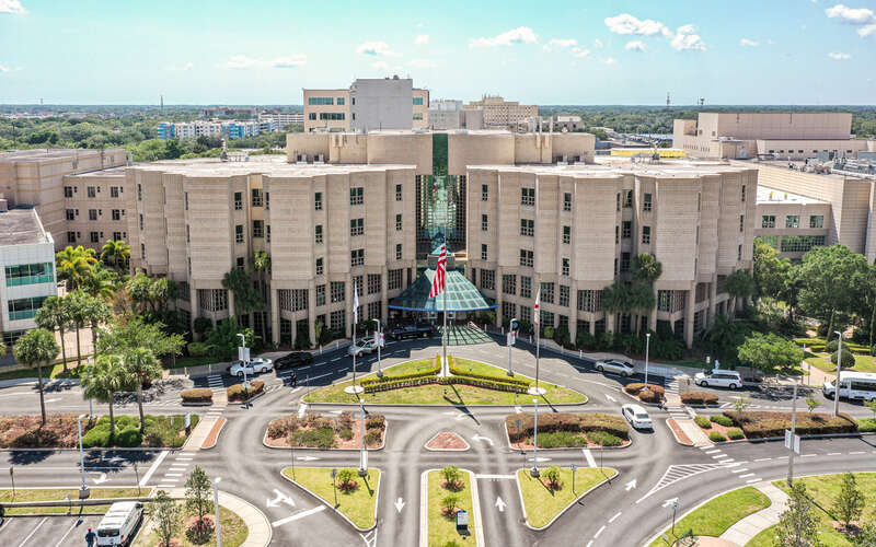
#1 Cancer Hospital in Tampa
Moffitt is the top-ranked cancer hospital in Tampa and central Florida and ranked No. 16 in the nation in Newsweek’s “America’s Best Cancer Hospitals."
Frequently asked questions (FAQs) about rectal cancer diagnosis
Because no single test provides every piece of information needed to plan rectal cancer treatment, the patient may undergo several examinations after an abnormality is first identified. Understanding the purpose of each test can make the diagnostic and staging process easier to follow.
What is the most accurate test used to confirm a rectal cancer diagnosis?
A tissue biopsy is the definitive test for confirming rectal cancer. Tissue is usually collected during a colonoscopy or sigmoidoscopy and examined under a microscope by a pathologist to determine whether cancer cells are present.
How long does it take to get biopsy results after a colonoscopy or sigmoidoscopy?
The timing varies depending on the laboratory and whether additional studies are required. Initial pathology results are often available within several business days, while specialized stains, biomarker testing or molecular profiling can require additional time.
Are there specific blood tests or tumor markers that can detect rectal cancer?
There is no blood test that can reliably diagnose rectal cancer by itself. CEA is the principal tumor marker used in rectal cancer care, but the CEA test lacks the sensitivity and specificity needed for screening or diagnosis and is primarily useful after cancer has been diagnosed.
Why is a pelvic MRI usually required after a rectal cancer diagnosis?
A high-resolution rectal protocol pelvic MRI provides detailed information about how far the tumor extends through the rectal wall, whether nearby lymph nodes appear to be involved and how close the cancer is to structures such as the mesorectal fascia and sphincter complex. These findings are important for assigning a clinical stage and determining the most appropriate sequence of chemotherapy, radiation therapy and surgery.
Does a biopsy tell what stage the rectal cancer is?
Not on its own. A biopsy confirms a diagnosis of rectal cancer and provides information about the cancer’s microscopic characteristics, while imaging and other clinical findings show how deeply the tumor has grown and whether it has reached lymph nodes or distant organs. The results are combined to determine the clinical stage before treatment.
Is endoscopic ultrasound required for every patient with rectal cancer?
No. A rectal protocol pelvic MRI is generally preferred for local staging. Endoscopic ultrasound can provide valuable information in selected cases, particularly when the healthcare team needs to determine whether an early tumor is T1, meaning it has grown into the layer of tissue beneath the inner lining of the rectum, or T2, meaning it has grown into the muscle layer of the rectal wall. It may also be used when MRI cannot be performed.
The Moffitt Cancer Center difference for diagnosing rectal cancer
An accurate rectal cancer diagnosis requires more than confirming that a tumor is malignant. Its exact location, depth, relationship to surrounding structures, pathological characteristics and molecular profile can all influence treatment. Moffitt’s Gastrointestinal Oncology Program brings these components together so the patient’s diagnostic findings can be evaluated as a whole.
Moffitt has specialists dedicated to rectal cancer, including surgical oncologists, medical oncologists, radiation oncologists, radiologists and pathologists. Each new patient’s case is reviewed by a multispecialty team, allowing experts with different areas of expertise to collectively evaluate the diagnostic findings and develop an individualized treatment plan.
Patients who have already received a diagnosis elsewhere can also turn to Moffitt for a second opinion. Subspecialized pathologists can review biopsy tissue to verify the diagnosis and characterize the tumor, while specialized radiologists can evaluate imaging to clarify the cancer’s location and extent. When appropriate, additional biomarker or molecular testing can provide further information about the tumor’s biology and potential treatment options.
Moffitt also offers advanced diagnostic capabilities within a comprehensive cancer center, including specialized gastrointestinal endoscopy, MRI and other imaging services. Our Endoscopic Oncology Program provides endoscopic ultrasonography for locoregional staging of gastrointestinal cancers, including rectal cancer.
To learn more about diagnosing rectal cancer, you can request an appointment with a specialist in Moffitt’s Gastrointestinal Oncology Program by calling 1-888-663-3488 or submitting a new patient registration form online. We do not require referrals.
Diagnosis