Mammogram Testing for Breast Cancer
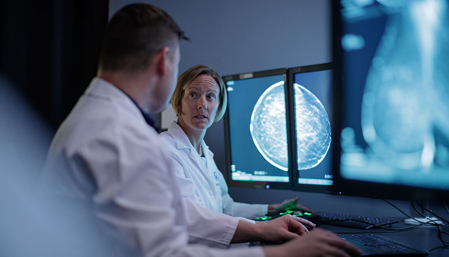
A mammogram is an imaging technique used for the early detection of breast cancer. After compressing the breast, a mammography technologist will use a low-dose X-ray system to capture highly detailed images of the breast tissue. The resulting images will be interpreted by a radiologist who can identify lumps, masses, calcifications and other unusual changes that may otherwise be unnoticeable.
An important component of a breast health strategy, routine screening mammograms can detect breast cancer at an early stage when there are generally more treatment options available. This helps many women achieve the best possible outcome and quality of life through timely intervention and management. The multispecialty team in the Don & Erika Wallace Comprehensive Breast Program at Moffitt Cancer Center recommends annual screening mammograms beginning at age 40 for most women, and earlier for those who have a heightened risk of developing breast cancer due to their family history or other factors.
Types of mammograms
In addition to screening for breast cancer, mammograms play an essential role in the diagnostic process. A physician may order a diagnostic mammogram to evaluate possible breast cancer symptoms found during a breast self-exam or a clinical breast exam, such as a lump or change in the size, shape, volume, texture or skin of a breast.
Moffitt offers digital breast tomosynthesis (3D mammography) for both screening and diagnostic purposes. This advanced breast imaging technique captures multiple X-rays from different angles, which are then reconstructed by a computer to create a highly detailed, three-dimensional depiction of the breast tissue. Compared to standard, two-dimensional mammography, 3D mammography produces images with better clarity, making it easier for a radiologist to detect abnormalities, particularly in women with dense breast tissue.
The Right Diagnosis. Right Away.
If you've received an abnormal test result that could indicate breast cancer, request an appointment with our Breast Oncology team today. Moffitt's diagnostic experts will perform the tests needed to diagnose or rule out cancer so you can know for sure.
Moffitt has the highest quality imaging technology and uses the least invasive testing procedures to give you accurate results.
What are dense breasts?
When discussing the results of a mammogram with a patient, the physician may tell her that her breasts are low- or high-density. Breast density refers to the relative proportion of different types of tissue in the breast, such as:
- Fatty tissue – Gives the breast its shape and size
- Glandular tissue – Produces breastmilk
- Fibrous tissue – Holds fatty and glandular breast tissue in place
Based on the appearance of the breast tissue in mammogram images, a radiologist will determine the patient’s breast density based on the Breast Imaging Reporting and Database System (BI-RADS), which is commonly used to report mammogram results. The BI-RADS classifies breast tissue into one or four categories of density:
- Almost entirely fatty – The breast primarily consists of fatty tissue.
- Scattered density – The breast is largely made up of fatty tissue with a few small pockets of fibrous and glandular tissue.
- Heterogeneously dense – The breast has many areas of fibrous and glandular tissue and relatively few areas of fatty tissue.
- Extremely dense – The breast is mostly composed of fibrous and glandular tissue and has minimal fatty tissue.
Physicians use the term “dense breasts” when referring to breasts that have a higher proportion of fibrous and glandular (nonfatty) tissue than fatty tissue. Dense breasts are very common, especially among young women. The classification is significant because fatty breast tissue looks dark in mammogram images, while both dense breast tissue and cancerous breast tissue appear white. Therefore, dense breast tissue can potentially conceal abnormalities in a mammogram, making it more difficult for a radiologist to detect breast lumps and tumors.
Mammograms for women with dense breasts
A physician may advise a woman with dense breasts to have more frequent screening mammograms along with additional testing, such as periodic breast ultrasounds or magnetic resonance imaging (MRI) scans. The physician may also recommend 3D mammography, which can allow a radiologist to view the breast tissue more clearly beyond the areas of density.
Another possible option is molecular breast imaging (MBI), a specialized, FDA-approved imaging technique that involves the use of a radioactive tracer that will accumulate in abnormal breast tissue, which will highlight any abnormalities in the resulting images. MBI can be particularly useful for identifying small tumors and assessing the breast health of women with dense breast tissue.
Mammograms for women with breast implants
The most common types of breast implants—silicone and saline—are widely considered to be safe and do not increase a woman’s risk of developing breast cancer. However, implants can conceal abnormalities in mammogram images.
Because it can be challenging for a mammography technologist to capture clear images of the breast tissue behind an implant, the procedure requires specialized expertise. In addition, the technologist should be informed in advance that they will be examining a patient who has breast implants because multiple images (implant displacement views) are usually needed. To capture the implant displacement views, the technologist will push the implant toward the chest wall and pull the breast tissue forward. Mammography presents a small risk of implant rupture.
What if my mammogram shows an abnormality?
A change in breast tissue detected in a mammogram image does not conclusively indicate cancer. In fact, most abnormalities found through mammography are ultimately determined to be benign. To make this determination, a physician will typically order additional testing, such as a breast ultrasound or magnetic resonance imaging (MRI) scan, which can provide more detailed images. If further testing is inconclusive, the physician may order a biopsy, which involves removing a small sample of suspicious tissue for microscopic evaluation by a pathologist, who can identify cancer cells.
Mammogram locations at Moffitt
Moffitt offers mammograms at two convenient locations:
Richard M. Schulze Family Foundation Outpatient Center at McKinley Campus
10920 N. McKinley Drive, Tampa, FL 33612
Monday, Wednesday, Thursday and Friday | 7:15 a.m. to 5:30 p.m.
Tuesday | 7:15 a.m. to 7 p.m.
Moffitt Cancer Center at International Plaza
4101 Jim Walter Blvd., Tampa, FL 33607
Monday and Thursday | 7 a.m. to 12 p.m.
Tuesday, Wednesday & Friday | 7 a.m. to 3:30 p.m.
Breast cancer risk assessments at Moffitt
At Moffitt, we encourage all women to learn about the risk factors for breast cancer so they can take action to prevent it or detect it early when it is easier to treat. Moffitt stands out from many other cancer screening providers because we evaluate breast cancer risk factors for every patient who comes to us for screening. This helps us identify women who have an elevated risk for breast cancer and could benefit from more intensive screening beyond an annual mammogram.

Breast cancer risk factors that we assess
When a patient is screened for breast cancer at Moffitt, our specialists provide an individualized risk assessment along with guidance on how to manage the risk. Some of the many risk factors we evaluate include:
Family history
Women who have one or more close family members who were diagnosed with breast cancer face a significantly higher risk. However, both women and their healthcare providers tend to overestimate the importance of family history when calculating breast cancer risk. The vast majority of women diagnosed with breast cancer have no family history of the disease.
Inherited gene mutations
Certain inherited mutations in breast cancer gene 1 (BRCA1) and breast cancer gene 2 (BRCA2) can lead to rapid breast cell growth and division, increasing the risk of breast cancer.
Age
Breast cancer risk increases with age. Most new cases are diagnosed in women 50 and older, while approximately 9% of new cases are diagnosed in women younger than 45.
Reproductive history
Women who started menstruating before age 12 or experienced menopause after age 55 have a heightened risk of developing breast cancer due to their prolonged exposure to hormones.
Breast density
According to the American Cancer Society, women with dense breast tissue have a higher risk of developing breast cancer than women with fatty breast tissue. While the reasons are unclear, some experts believe dense breast tissue may have more cells that can undergo abnormal changes that lead to cancer.
Obesity
Carrying excess body weight, particularly after menopause, can increase a woman’s risk of developing breast cancer. Other factors commonly associated with obesity, such as poor nutrition and a sedentary lifestyle, can further increase the risk.
Prior radiation, drug or hormone therapy
Women who had radiation therapy delivered to their chest before the age of 30, took certain medications or used hormone therapy (including certain birth control pills) may have a higher breast cancer risk.
Breast cancer risk assessment clinics
In addition to providing individual risk assessments for breast cancer screening patients, Moffitt has established several specialized risk assessment clinics. These include our:
- Breast Evaluation Clinic – For patients who have breast cancer symptoms or an abnormal imaging study performed at another facility
- Breast Surveillance Clinic – For women at heightened risk of developing breast cancer due to a medical condition
- Breast Cancer Survivorship Clinic – A free, eight-week workshop designed to help breast cancer survivors transition from active treatment, including strategies for proper nutrition, stress management and exercise
- Familial Breast and Ovarian Cancer Screening and Prevention Clinic – Provides comprehensive care and counseling for women at increased risk of developing breast and/or ovarian cancer due to their family history, including appropriate surveillance testing recommendations, such as mammograms, transvaginal ultrasounds, Pap smears, breast MRIs and CA-125 blood tests
Diagnosis matters. Choose Moffitt first.
The subspecialized breast imaging radiologists at Moffitt are highly skilled and experienced in screening and diagnosing breast cancer, particularly in women with dense breast tissue or breast implants. Our comprehensive screening services are complemented by individualized advice, breakthrough treatments and compassionate support from a multispecialty team that focuses exclusively on breast cancer.
If you would like to request an appointment for a mammogram and risk assessment at Moffitt—Florida’s No. 1 cancer hospital—call 1-888-663-3488 or submit a new patient registration form online. Moffitt is streamlining access to world-class cancer care by welcoming new patients without referrals.
References
American Cancer Society: Mammograms for Women With Breast Implants
Helpful Links:
Screening
