Inflammatory Breast Cancer Diagnosis
Inflammatory breast cancer (IBC) is a rare but aggressive type of breast cancer that occurs when clusters of abnormal cells form beneath the skin and block the lymphatic vessels within the breast. The blockage disrupts normal lymph drainage, leading to fluid buildup. Unlike most breast cancers, IBC typically does not present with a distinct lump. Instead, it may cause rapid changes in the appearance and texture of the breast, such as redness, swelling, warmth, skin thickening and pitting that resembles an orange peel (peau d’orange). Because these symptoms can mimic those of a breast infection or injury, the diagnosis is sometimes delayed.
Although inflammatory breast cancer can develop at any age, it is most frequently seen in women younger than 60. Because the cancer tends to progress quickly, early detection and prompt treatment are essential to achieve the best possible outcome and quality of life. Typically, the diagnostic process involves a physical examination, imaging studies and a biopsy.
How is inflammatory breast cancer detected?
In many cases, IBC is not found through a routine breast cancer screening test, mainly because it does not present as a distinct lump that can be felt during a clinical breast exam or seen on a mammogram. Instead, the cancer is often identified after its hallmark symptoms—peau d’orange and other breast skin changes—prompt a medical evaluation.
Imaging tests used for diagnosing inflammatory breast cancer
When IBC is suspected based on physical symptoms, imaging usually plays a key role in the diagnostic process, allowing the physician to evaluate subtle changes in the breast tissue and identify signs that might not be apparent during a physical exam. It can also help the physician assess the extent of the cancer and determine whether it has spread to nearby lymph nodes or surrounding structures. Common imaging tests used to evaluate IBC include:
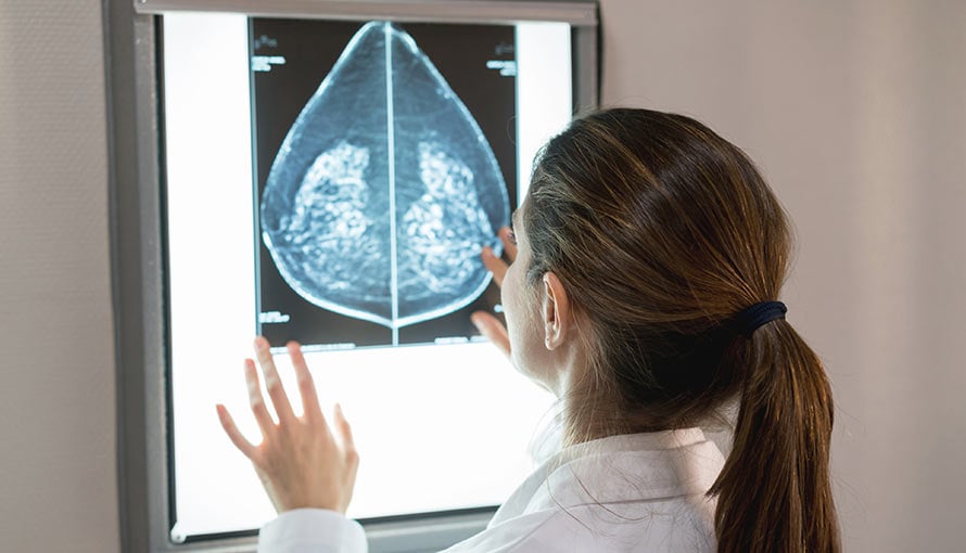
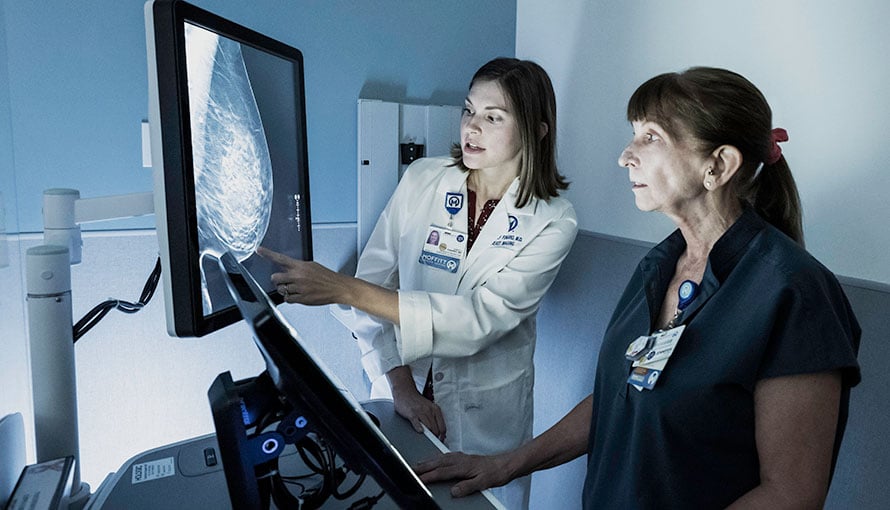
Diagnostic mammogram
Mammography is often the first imaging test ordered when inflammatory breast cancer is suspected. Using low-dose X-rays, this test produces detailed images of the breast tissue. Although a mammogram is unlikely to show a distinct lump in a case of IBC, it might reveal thickened skin, increased breast density or enlarged lymph nodes, which may warrant further evaluation. Mammography also allows the physician to compare both breasts and identify any asymmetric changes that could support the diagnosis.

"Although we understood the severity of what I was dealing with, Dr. Susan Hoover promised and assured me that I was going to be on the other side of cancer in two years."
Tiffany, Breast Cancer Survivor
Request an AppointmentBreast ultrasound
Breast ultrasound uses high-frequency sound waves to create real-time visuals of the breast tissue and surrounding structures. This imaging test can be particularly helpful for evaluating thickened skin, fluid buildup and changes in the underlying tissue layers. Ultrasound can also be used to guide a needle biopsy or help the physician assess whether nearby lymph nodes are enlarged or otherwise abnormal.
Breast magnetic resonance imaging (MRI)
Breast MRI is often recommended when mammography or ultrasound findings are inconclusive or when more detailed information is needed to guide a biopsy or develop a treatment plan. This advanced, noninvasive imaging technique provides high-resolution images that can help the physician evaluate the full extent of inflammatory breast cancer, including any potential involvement of the skin, lymph nodes and chest wall. During the procedure, the patient lies on a specially designed table that slides into a cylindrical scanner, where magnetic fields and radio waves generate cross-sectional images of the breast and surrounding tissues.
Computed tomography (CT) scan of the chest, abdomen and pelvis
After confirming a diagnosis of inflammatory breast cancer, the physician may order a CT scan to stage the cancer and evaluate whether it has spread beyond the breast and regional lymph nodes. During the test, an X-ray beam will move in a circular pattern around the patient’s body, capturing multiple images of the same internal structures from multiple angles. The X-ray data will then be processed by a computer to create layered images of the lungs, liver and other organs, which may reveal evidence of cancer spread.
Positron emission tomography (PET) scan
A PET scan, often combined with a CT scan (PET/CT), can help the physician detect areas of high metabolic activity within the body, which may suggest cancer spread. This advanced nuclear imaging test can provide a comprehensive overview of the cancer’s reach, which is important for treatment planning. It can be particularly valuable in assessing distant metastasis of an aggressive cancer, such as IBC.
Breast Cancer Outcomes
The goal of any cancer treatment is to achieve the most favorable outcome while minimizing side effects and ensuring the best possible quality of life. On average, Moffitt's breast cancer treatment survival rates are 1.5 times the national average.
Procedures used for diagnosing inflammatory breast cancer
When IBC is suspected, imaging tests are often followed by one or more procedures to confirm the diagnosis. Because this type of cancer tends to grow rapidly and does not usually present as a distinct lump, tissue sampling and laboratory analyses are essential to detect cancerous cells, determine the cancer subtype and guide treatment planning. Diagnostic procedures commonly used in the evaluation of IBC include:
Skin punch biopsy
A skin punch biopsy involves the use of a specialized tool to remove a small, circular sample of skin tissue. Because inflammatory breast cancer often affects the skin by causing redness, swelling and thickening, this type of biopsy can help the physician confirm the presence of cancerous cells in the dermal lymphatic vessels. This relatively quick procedure is typically performed under local anesthesia in an outpatient setting.
Core needle biopsy
Core needle biopsy is the standard technique for obtaining tissue samples from within the breast. Using a hollow needle, the physician will remove small cylinders of tissue from an area of concern identified through diagnostic imaging. The physician may use mammography or ultrasound imaging to precisely guide the needle into position. The collected tissue will then be sent to a laboratory, where a pathologist will examine the samples under a microscope to detect cancerous cells and assess their specific characteristics.
Fine-needle aspiration (FNA)
Fine-needle aspiration can be used to sample tissue or fluid from a suspicious lymph node or mass. After inserting a thin, hollow needle into the targeted area, the physician will extract a small number of cells. While FNA provides less tissue for testing than core needle biopsy, the results can help the physician determine whether IBC has invaded nearby lymph nodes.
Sentinel lymph node biopsy or lymph node sampling
Lymph node evaluation is an important step in staging inflammatory breast cancer. If a physical examination or imaging study suggests lymph node involvement, the physician may perform a biopsy of the sentinel lymph node, which is the first node to which the cancer is most likely to spread. This test can be performed through fine-needle aspiration, core needle biopsy or surgery.
Laboratory tests used for diagnosing inflammatory breast cancer
Lab testing is essential for definitively diagnosing inflammatory breast cancer, as it allows for the microscopic evaluation of tissue samples obtained through biopsy. In addition to confirming the presence of cancer cells, certain lab tests can provide valuable molecular and systemic insights that support staging and treatment planning. These tests can help the physician assess the patient’s overall health, determine how far the cancer has progressed and identify biomarkers that may guide targeted therapies. Common laboratory tests used in these contexts include:
Pathology testing of biopsy samples
After a small amount of tissue is taken through a skin punch biopsy, core needle biopsy, FNA or surgical procedure, the sample will be sent to a pathology lab for detailed analysis. This is essential for confirming the presence of cancer cells and determining their characteristics, such as:
- Hormone receptor status (ER/PR)
- Human epidermal growth factor receptor 2 (HER2) status
- Cancer grade
The results of pathology testing can help the physician classify the cancer and determine the most effective treatment approach.
Blood work
While not specifically used for diagnosing inflammatory breast cancer, certain blood tests may be used to support the physician’s overall assessment and detect signs of metastasis. These include:
- Complete blood count (CBC) – Evaluates the levels of red and white blood cells and platelets in the bloodstream, which may be affected by cancer or its treatment
- Liver function test (LFT) – Assesses the health of the liver and may reveal signs of cancer spread
- Alkaline phosphatase (ALP) and lactate dehydrogenase (LDH) – Checks for elevated levels of ALP or LDH, which may suggest bone or liver involvement
Tumor marker testing
In select cases, the physician may measure tumor markers, such as CA 15-3 or CEA, to monitor the patient’s response to treatment or detect cancer recurrence. However, tumor marker testing is not specific or reliable enough to confirm an initial diagnosis of IBC.
Benefit from world-class care at Moffitt Cancer Center
Inflammatory breast cancer is relatively uncommon. It is also a fast-growing cancer that requires highly specialized and aggressive treatment. Moffitt is home to the Don & Erika Wallace Comprehensive Breast Program, a comprehensive, high-volume breast clinic that offers our patients access to vast expertise and resources, including board-certified physicians and a robust portfolio of clinical trials.
If you would like to learn more about the diagnostic process for inflammatory breast cancer, you can request an appointment with a specialist at Moffitt by calling 1-888-663-3488 or submitting a new patient registration form online. We do not require referrals.
Diagnosis