A Closer Look into the Future
Key Takeaways:
- Moffitt Cancer Center is digitizing up to 95% of pathology slides to speed diagnoses and advance personalized cancer care
- Digital scanners can turn glass slides into high-resolution images in four to five minutes, replacing processes that once took days or weeks
- Artificial intelligence could help pathologists detect subtle cancer patterns, guide treatment decisions and develop new approaches for rare cancers
Long before a patient hears the words “it’s cancer,” a pathologist has already searched for the answer beneath the microscope.
Under its lens, scattered cells become stories, silent tissues reveal hidden disease, and answers emerge from details too small to see with the naked eye. The instrument is far more than glass and light — it becomes a pathologist’s closest partner.
“The identity of the pathologist is tied to the microscope,” said William Westra, MD, vice chair of Anatomic Pathology at Moffitt Cancer Center.
So, when Westra was told he may not need his microscope anymore after relying on it for decades, he was resistant.
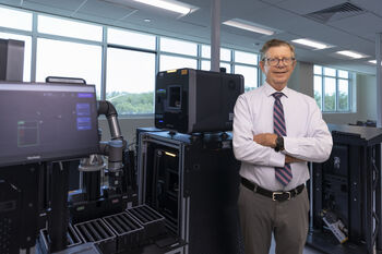
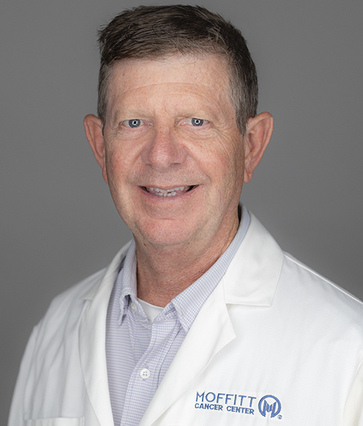
William Westra, MD, is spearheading the shift to digital pathology, which will improve diagnostic capabilities and help patients get answers faster.
He had been tasked with ushering in the next generation of disease study called digital pathology. Using large digital scanners instead of a microscope, pathologists can transfer histologic images into high-resolution digital images.
“You might think, well, what’s the big deal about that? You’re still looking at the same images, but once they’re on a computer monitor, you now have this computational platform where you can do all kinds of things,” Westra said.
It became a good enough reason for Westra to ditch his microscope and become digital pathology’s “biggest cheerleader,” as he puts it.
With a digital platform, pathologists can send and receive images from around the world for second opinions and consultations. They can also use artificial intelligence to help make more accurate diagnoses through clinical diagnostic assistance, expedite the triage of patient cases and match patients to specific treatments.
Moffitt has become a leader in the digital pathology space, operating as one of the only major cancer centers to commence the full adoption of the system across the entire institution. The cancer center is currently working to digitize 90% to 95% of all slides to help open the door even further into personalized medicine for patients.
It Starts With Digital Scanners
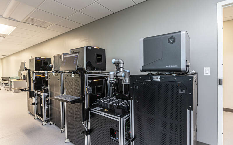
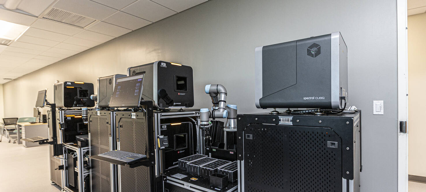
Moffitt’s digital pathology lab currently has nine digital scanners that can convert traditional glass slides into high-resolution images in just minutes by using high throughput technologies.
The machines resemble robots. An automated arm picks up a slide and places it inside the scanner, where a built-in microscope creates a digital image that appears on the scanner’s computer monitor. Those images are stored in a digital cloud where they can be accessed by Moffitt pathologists, medical oncologists, surgeons and scientists, as well as shared with other clinicians around the world with just the click of a button. The arm then removes the slide and places it in a storage area for archiving before picking up a new slide to be scanned.
Moffitt partnered with the state this year to secure $2 million to launch the program and will receive an additional $3.7 million over the next year. Those funds are combined with about $15 million pooled together from hundreds of individual donations to Moffitt’s Foundation.
The cancer center will generate an estimated 1.7 million glass slides from patient specimens in the lab’s first year, with the number of slides increasing 7% to 10% each year.
The digital cloud can currently hold 24 petabytes of data. A petabyte is 1 million gigabytes, and just one petabyte could hold about 250 million photos on a smartphone or store around 1 billion e-books.
-

1.7 Million
Number of glass slides generated from patient specimens in the lab’s first year
-

24
Number of petabytes of data the digital cloud can hold
With such a high slide volume, Moffitt’s initiative to digitize them will be a major time-saver. Traditionally, slides require strict packaging, labeling and careful transportation. Now, they can be analyzed by the scanner in four to five minutes and viewed and shared by pathologists almost instantly.
“From that traditional glass slide practice to the digital platform, we will reduce turnaround time from days and weeks to minutes to hours without losing a single slide,” Westra said.
A shorter turnaround benefits patients immensely. A quicker diagnosis means patients get answers faster and can start treatment sooner.
The digital platform also solves practical issues, like the projected shortage of pathologists nationwide over the next few decades while case volumes continue to rise.
“We have begun to use digital pathologists who can work at remote locations, which lets us be more efficient in the demands moving forward,” Westra said.
Sharing information remotely also puts Moffitt in a position to expand its reach to more rural and underserved areas across the state that may have limited diagnostic expertise. Instead of transporting a slide hundreds of miles, the digitized image can quickly and easily be used during a virtual consultation.
“We could provide these healthcare institutions with a digital scanner and train them how to use it, and the images could be sent over digitally and read by pathologists here,” Westra said.
Using AI Algorithms
One of the most important jobs a pathologist has is determining case diagnoses and prognoses. While that has been done with a microscope and the pathologist’s naked eye in the past, digital pathology will use artificial intelligence to increase workflow efficiency and diagnostic accuracy, measure biomarkers, and help predict treatment response.
AI can identify which cases are more advanced and would benefit from earlier treatment, placing those cases at the top of a pathologist’s queue to review. The system can also flag suspicious areas on slides, helping pathologists focus attention on the most important regions first.
Moffitt is now selecting AI algorithms for specific cancer types that can identify subtle patterns that may be more difficult for the human eye to detect, reducing the risk of missed diagnoses and helping standardize interpretations between pathologists. These algorithms can also measure predictive biomarkers — such as HER2 in breast cancer — more consistently, which can help guide treatment decisions.
“Application of the AI-assisted algorithms will allow us to have faster turnaround time and more accurate diagnoses,” Westra said.
'Ultimate Level of Information'
Ignacio Wistuba, MD, has also had a front-row seat to the shift away from the microscope. He says he had zero hesitation to embrace the change.
“I am very different. I never liked the microscope,” explained Wistuba, chair of Moffitt’s Translational Pathology Department. “It always felt to me like a black box. Now there is a way to get a more complete and detailed record of what one is looking at.”
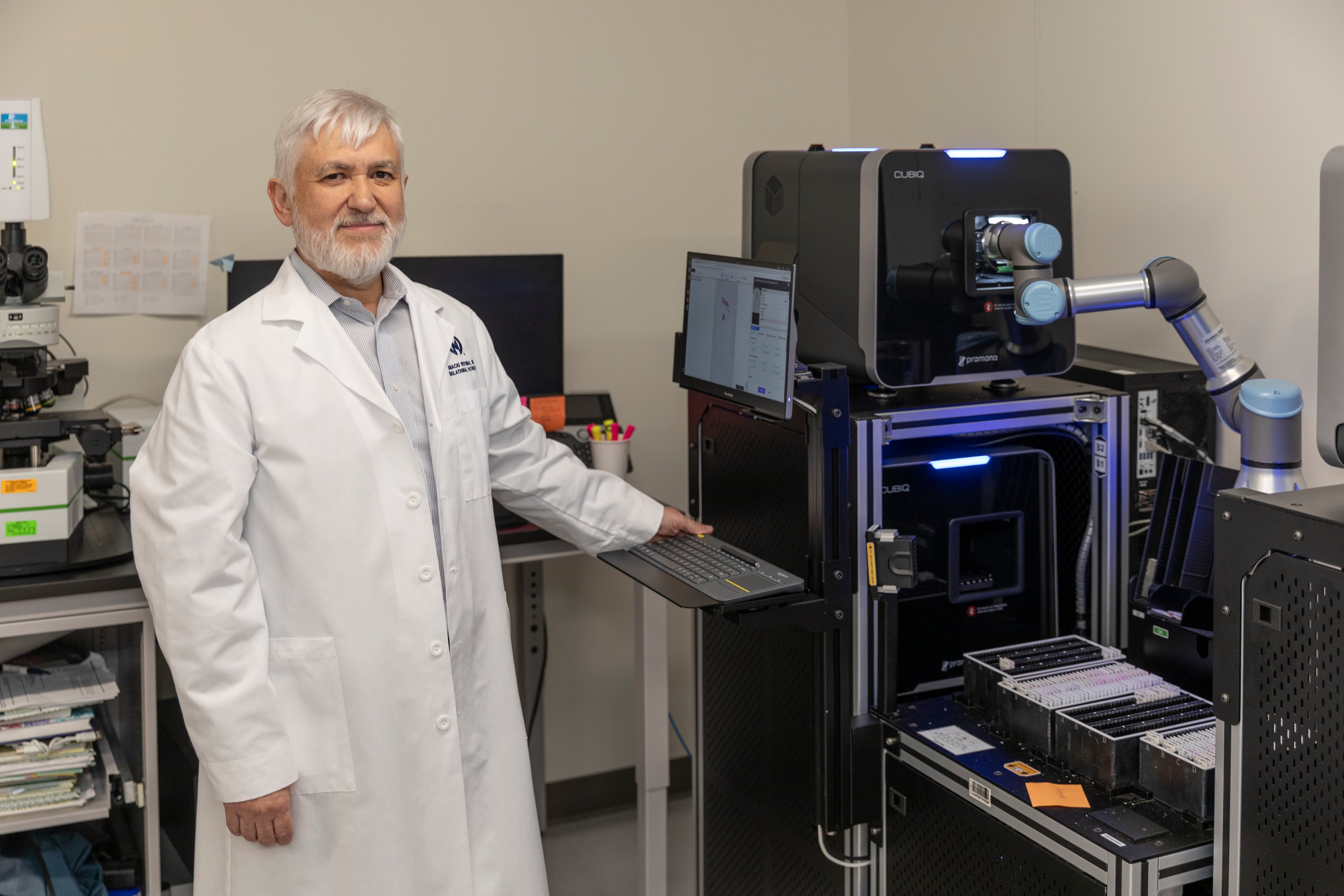
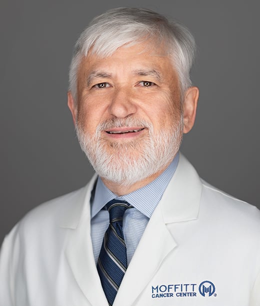
Ignacio Wistuba, MD, is chair of Moffitt’s new Translational Pathology Department, which focuses on discovering the origin of cancer and identifying biomarkers to target.
Stepping away from his black box and into the digital world has radically changed research capabilities. While about 95% of pathology is dedicated to diagnosing disease, Wistuba says he lives in the other 5%, which is a group of translational pathologists that focuses on discovering the origin of cancer and identifying biomarkers to target.
Translational pathology aims to merge the work between clinicians at the bedside and researchers aiming to find new treatments.
“How can we use human specimens to see what we can learn? For example, we can develop a predictive biomarker — something to tell us how a patient will respond — or a mechanism to improve responses or prevent treatment resistance,” Wistuba said.
While Moffitt is already doing molecular and genomic studies on tumor samples, many answers still may lie in the tissue, such as the architecture and patterns in cells, that can only be analyzed using digital images.
“We can interrogate them to see if they can give us that information that is somehow hidden,” Wistuba said. “By running analyses on the tissue, we can see what we can learn from them and use in the future. That is what I would call the ultimate level of information.”
This work can be applied in clinical trials testing new targeted therapies or combination treatments. Using biopsies and blood tests before and during treatment, translational pathologists can monitor biomarkers on a regular basis to investigate how the patient is responding to treatment. This can help identify early signs of treatment success or failure, which can be used to create predictive biomarkers to help other patients in the future.
Building AI for Rare Cancers
AI also plays a major role in the translational pathology team’s research. The tool will help integrate information from the digital scans with other clinical and molecular data for a large number of cases.
“We will learn from thousands of patients and then develop predictive algorithms,” Wistuba said. “A clinician can see a patient and based on the features of that particular case, predict that the chances of being successful with this treatment is X, or that there are three options and you will respond best to Y treatment.”
Using AI to analyze more commonly diagnosed cancers, like breast and prostate cancer, is easier, and there are already AI-assisted algorithms available to help investigate those samples.
Wistuba and his team are setting their sights on rarer cancers, such as neuroendocrine tumors and sarcoma. They are working to develop their own AI algorithms to assist in analyzing digital scans for these rarer diseases more often seen at Moffitt.
“My goal is to start doing clinical trials that are driven by translational studies that can take us to the next level, which is biomarker-driven clinical trials that are novel and can truly change outcomes,” Wistuba said. “And then we plan to take those models and apply them to rare cancers.”
'One-Stop Shopping'
Both Westra and Wistuba have spent their careers witnessing the evolution of pathology — from the first sequencing to spatial analyses to now computational and digital pathology. By instituting the Moffitt-wide digitization of slides, pathologists have what Wistuba calls “one-stop shopping,” the ability to analyze samples at every single level.
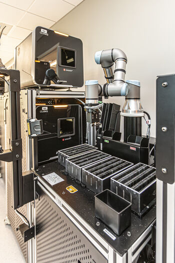
Moffitt's digital scanners can convert traditional glass slides into high-resolution images in just minutes. The cancer center now plans on purchasing additional scanners and storage space.
Moffitt is already focused on the growth of the program. The team plans to add four more digital scanners in the next year to meet increased demand, with one additional scanner for the next five years. Starting at year 6, two new scanners will need to be purchased each year.
Moffitt will also eventually need more digital storage space. The 24-petabyte cloud is expected to be at capacity in about 2 ½ years, and the program will have to continually update the scanners’ software as technology advances over the years.
The future also holds opportunities to use the digital platform for cytology, which focuses on examining individual cells or small clusters of cells to screen for, diagnose or monitor cancer. Those slides will require a three-dimentional scan that will take more time to process and more space to store.
It’s a future that will change patient care and outcomes, and one that most likely will not involve a favorite tool of the past: the microscope.
 Medically reviewed by William Westra, MD, Anatomic Pathology Department