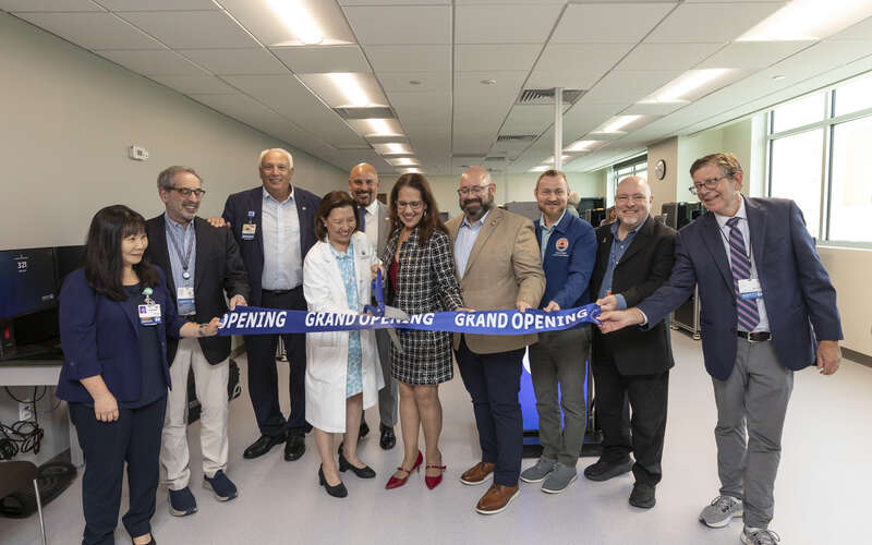
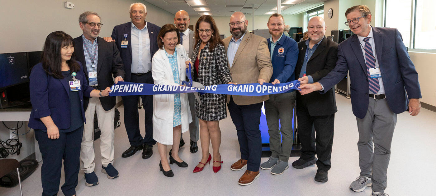
Moffitt Unveils New Digital Pathology Lab
Key Takeaways:
• Moffitt’s new Digital Pathology Lab will digitize up to 95% of slides
• Digital scanners cut pathology consultation turnaround times from days or weeks to hours
• AI tools can improve diagnostic accuracy, prioritize urgent cases and identify biomarkers for treatment decisions
Stepping into the new Digital Pathology Lab at Moffitt Cancer Center is like stepping into the future. The room is lined with digital scanners that will convert traditional glass microscope slides into high-resolution images in just minutes.
Once the slides are ready to be digitized, a robotic arm picks up a slide and places it inside the scanner. There, built-in microscopes create a digital image that appears on the scanner’s computer monitor. The robotic arm removes the slide and places it in a storage area for archiving before picking up a new slide to be scanned. This takes up to five minutes.
Those images will be stored in a digital cloud where they can be accessed by Moffitt pathologists and shared with other clinicians around the world. The application of artificial intelligence algorithms will allow pathologists to work faster and be more accurate with their diagnoses.
“The concept of digital pathology is very straightforward. We take histologic images and transfer those into high-resolution digital images,” said William Westra, MD, vice chair of Anatomic Pathology at Moffitt. “The ability to put those images on a computational platform really opens the door for endless novel applications.”
Reducing Turnaround Time
Moffitt currently has nine digital scanners paid for by state funding and money raised by Moffitt’s Foundation. The cancer center will generate an estimated 1.7 million glass slides from patient specimens in the 2026 fiscal year, with the number of slides increasing 7% to 10% each year. The goal is to digitize 90% to 95% of all slides and store the images in a cloud that can hold 24 petabytes of data. A petabyte is 1 million gigabytes, and just one petabyte can hold about 250 million photos on a smartphone or store around 1 billion e-books.
-

9
Digital scanners
-

1.7 million
Estimated glass slides from patient specimens in the 2026 fiscal year
-

90% to 95%
Of all slides will be digitized
-

24
Petabytes of data in digital cloud
Digitizing slides will be a major time-saver, especially when sending out cases to other pathologists for expert consultation. It enables pathologists to share images easily and instantly, compared to past practice that required strict packaging, labeling and transportation of slides through the mail delivery system.

“From that traditional glass slide practice to the digital platform we reduced turnaround time from days and weeks to minutes to hours without losing a single slide,” Westra said.
The system also provides an opportunity for Moffitt to expand its reach to rural areas by allowing for expert consults.
“These images can be shared in real time across institutions globally. The promise of digital pathology is to provide direct access to diagnosis expertise especially for underserved areas,” Westra said.
Harnessing Artificial Intelligence
Not only can pathologists use the high-resolution digital images to analyze specimens, but they can also harness the power of AI to improve workflow efficiency and diagnostic accuracy, measure biomarkers and help predict treatment response.
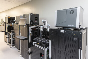
Moffitt has nine digital scanners with plans to add four more next year.
AI can identify which cases are more advanced and would benefit from earlier treatment, placing those cases at the top of a pathologist’s queue to review. The system can also flag suspicious areas on slides, helping pathologists focus attention on the most important regions first.
Moffitt is now selecting AI algorithms for specific cancer types that can identify subtle patterns that may be more difficult for the human eye to detect, reducing the risk of missed diagnoses and helping standardize interpretations between pathologists. These algorithms can also measure biomarkers — such as HER2 in breast cancer — more consistently, which can help guide treatment decisions.
Pathologists can also use the AI platforms to collaborate with other pathologists across the state and globe, discussing cases and inputting other data such as patient scans and history into the system to aid decision-making.
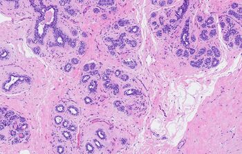
Once the slides are digitized, pathologists can use computational programs and AI to help triage patients, investigate biomarkers and match patients to treatments.
Most importantly, AI will enable the digital pathology team to work together with Moffitt’s Translational Pathology Department. The translational pathology team’s goal is to merge the work between clinicians at the bedside and the researchers working to find better treatments. Using the images generated by the digital pathology scanners, researchers can identify new biomarkers that can be targeted and investigate why some treatments fail.
“Application of the AI-assisted algorithms will allow us to have faster turnaround time and more accurate diagnoses,” Westra said. “And working with our translational pathology partners will help develop predictive algorithms and research that will benefit our patients.