Seeing Cancer Clearly: How Microscopes Are Transforming Care and Discovery
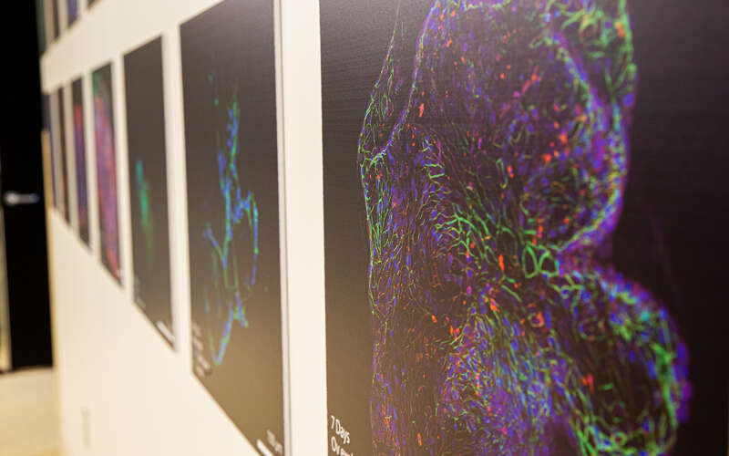
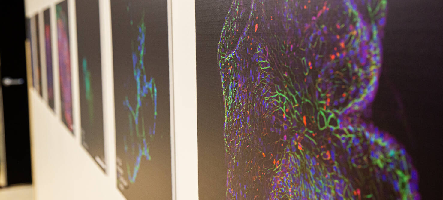
Each image tells a different story. A tumor under attack. Immune cells in motion. A therapy beginning to work. Together, they form what one researcher describes as a “periodic table of cancer,” capturing the diversity and complexity of the disease at the cellular level.
“Each one of these images represents a unique project, a disease site, an effort, a collaboration,” said Ryan Smolchek, PhD, a research bioengineer.
These images are more than scientific visuals. They are a powerful tool for understanding how cancer behaves and how it can be treated.
From Viewing to Experimenting
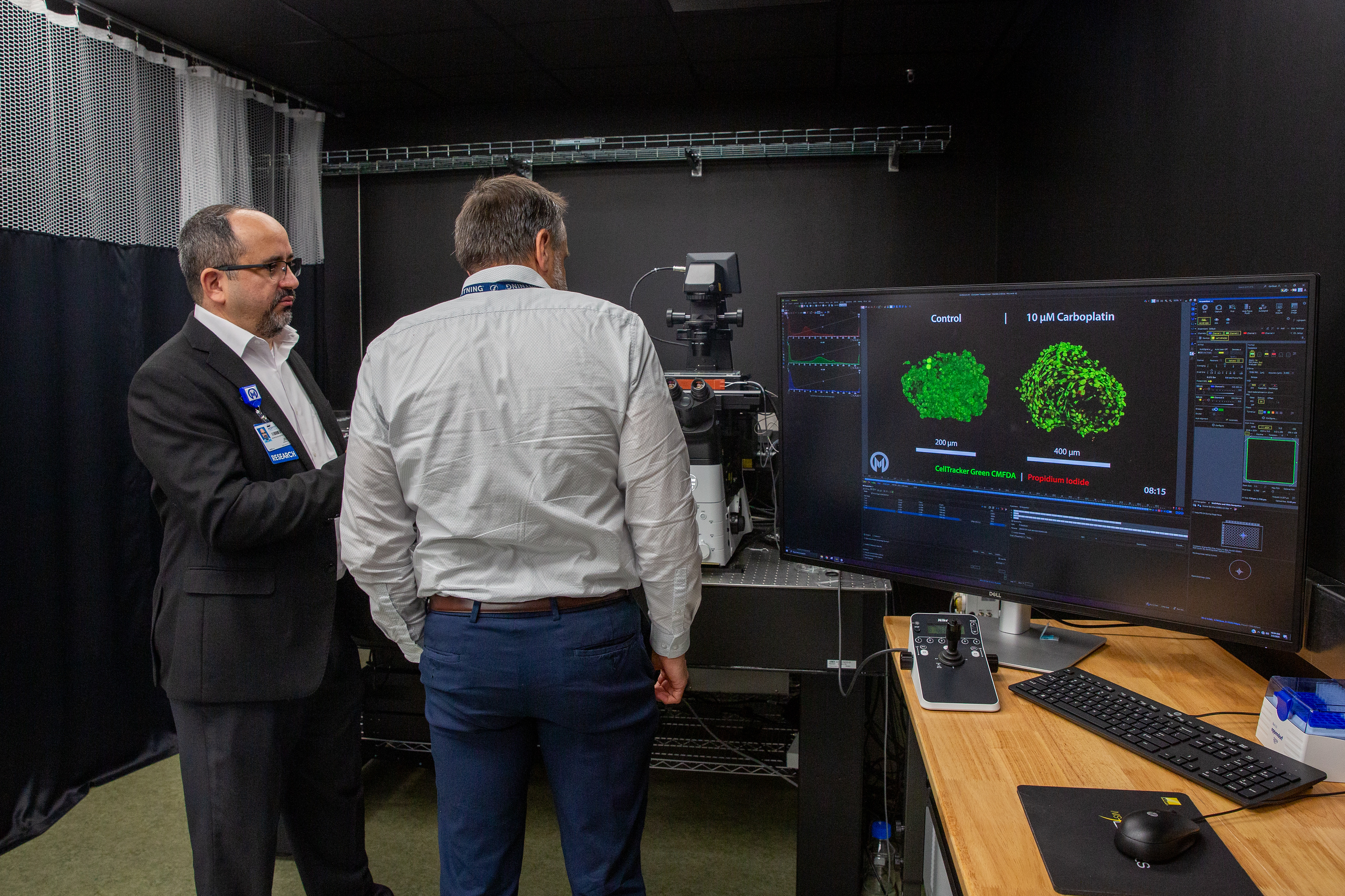
Jose Serrano-Velez, PhD, shows off an image captured in the Nikon Center of Excellence at Moffitt.
Microscopes have long played a role in diagnosing cancer, helping physicians examine tissue and identify disease. Today, their role has expanded far beyond observation.
“The microscopes are the tools that we use to visualize what’s going on with the cancer,” said Jose Serrano-Velez, PhD, Couch Bioimaging director of the Nikon Center of Excellence. “In the last 10 to 15 years, microscopes have been moving from taking images to actually performing experiments.”
Researchers can now place living tumor samples under advanced microscopes and observe what happens when therapies are introduced. Cells can be labeled to show whether they are alive or dying. Immune cells can be tracked as they move toward and interact with tumors.
Understanding How Treatments Work
This ability to observe cancer in real time is helping researchers answer critical questions.
In some experiments, scientists watch engineered immune cells attack tumors, capturing the moment cancer cells begin to break down. In others, they study how treatments such as bacteria-based therapies for bladder cancer reactivate immune cells that had previously been ineffective.
“We want to visualize it,” Smolchek said. “Is the bacteria being taken up by these cells? Are they presenting it?”
By answering these questions, researchers can better understand not only whether a treatment works, but why it works and how it might be improved.
Advancing Personalized Care
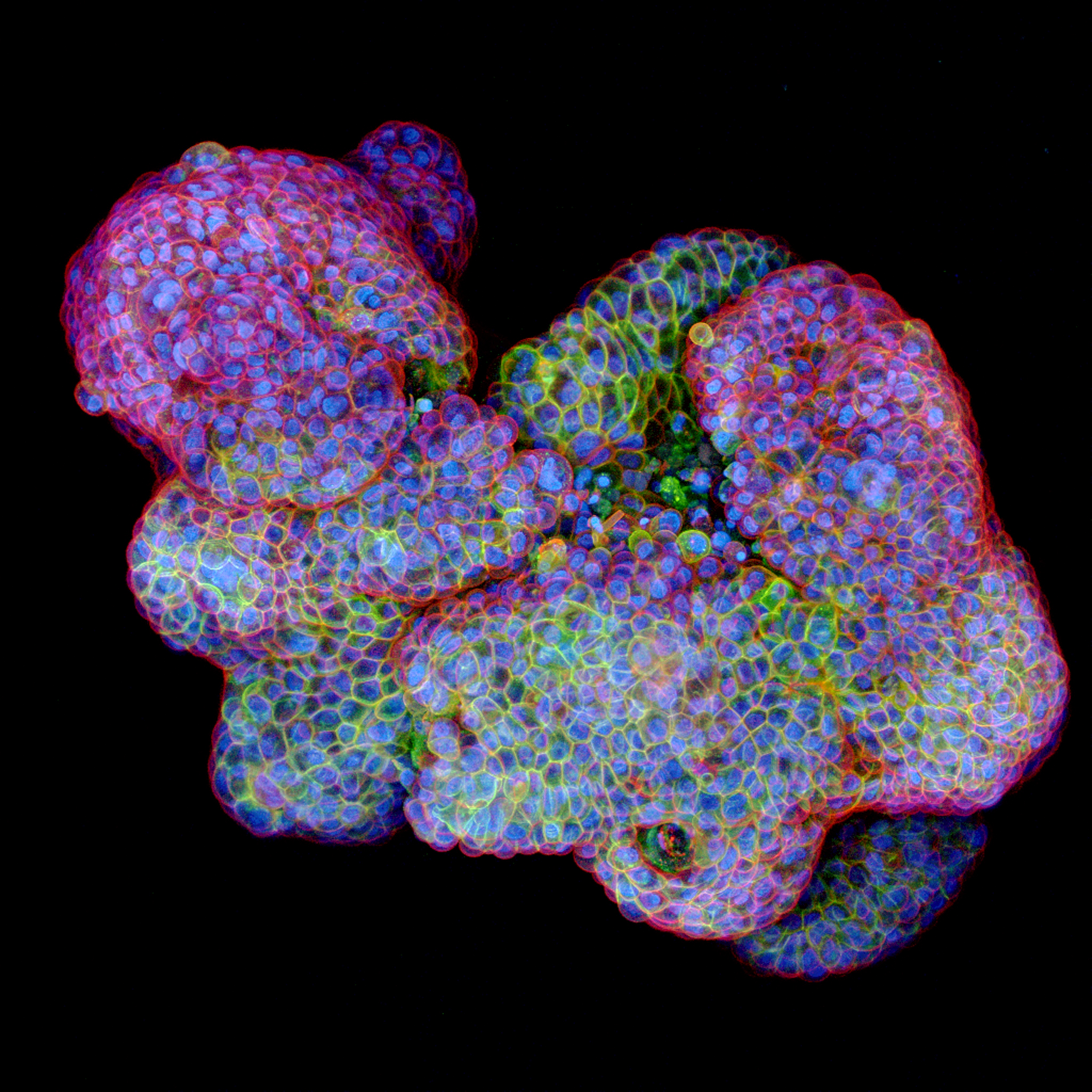
A colorful microscope image of cancer cells, where the bright green outlines show a protein on the cell surface that helps doctors determine if a targeted treatment is likely to work.
Microscopy is also helping move cancer care toward a more personalized approach.
Doctors often rely on genetic or molecular markers to guide treatment decisions. Microscopes offer a more direct way to evaluate how a patient’s tumor responds to different therapies.
“We can use microscopy to say, ‘This patient is overexpressing this protein, very likely to be a good candidate for this treatment,’” Smolchek said.
Researchers can also test multiple treatments on tumor samples and compare outcomes.
“You can actually say, ‘This one killed more of the tumor. That might be the better treatment option,’” he said.
This approach has the potential to reduce trial and error in treatment selection and provide faster, more precise care.
Capturing Cancer’s Complexity
Cancer is not a single type of cell. It is a complex system that includes tumor cells, immune cells and structural components that support growth.
Microscopes allow researchers to study these interactions in detail. Scientists can create three-dimensional models that preserve the structure and diversity of real tumors, including the presence of immune cells that are often missing from traditional models.
“We want to be able to keep the immune compartment,” Smolchek said.
This more complete view helps researchers better understand how tumors grow, how they evade the immune system and how they respond to treatment.
Technology and the Future
Advances in microscopy technology are driving rapid progress in cancer research.
Modern labs use a range of specialized microscopes that can capture fast-moving processes or reveal structures at extremely high resolution. These tools continue to improve, offering greater sensitivity and allowing researchers to study living cells with less disruption.
Looking ahead, scientists expect even more powerful capabilities.
“I think more high throughput and multiplexing, looking at different components of the cancer microenvironment at the same time,” Serrano-Velez said.
Artificial intelligence is also playing an increasing role. Researchers are using AI to analyze images, identify cells and process large datasets more efficiently.
“We are talking about thousands of samples, thousands of cells,” Serrano-Velez said.
A Clearer Path Forward
For researchers, microscopes provide a way to see cancer more clearly than ever before. For patients, that clarity could lead to better treatments and better outcomes.
“You can see how different efforts from different people get together to find a cure and prevent cancer,” Serrano-Velez said.
Each image captured under the microscope represents a step forward. And each step brings science closer to understanding, treating and ultimately overcoming cancer.