
AI-Driven Prostate Nomograms: How Moffitt Research Is Refining Focal Therapy Selection
Key Takeaways for Busy Providers:
- AI-driven prostate imaging tools are elevating nomograms by identifying where cancer sits in a gland, how much tissue it involves and how accurate that map is.
- AI systems for detecting clinically significant prostate cancer demonstrated higher accuracy and fewer false positives than experienced radiologist readings.
- Research demonstrates that AI-generated cancer maps predicted extracapsular extension better than established tools like Partin tables.
- Small, unifocal, MRI-visible lesions warrant a focal therapy conversation, and Moffitt’s GU oncology program can work through that decision alongside referring physicians.
- For appropriately selected patients, focal therapy is an outpatient procedure with minimal postoperative discomfort and a quicker recovery than radical prostatectomy, making accurate patient selection increasingly important.
Deciding which prostate cancer patients are good candidates for focal therapy has always come down to one hard question: not just whether the cancer is significant, but exactly where it sits, how much of the gland it touches and how much confidence a physician should place in that picture.
Traditional nomograms, built on population-level clinical and pathologic data, were never designed to answer that question with the precision focal therapy demands. A new generation of AI-enhanced imaging tools is starting to address that shortfall, and Moffitt Cancer Center's Genitourinary Oncology and Machine Learning teams are contributing to the research behind it.
Why Don't Traditional Nomograms Fit Focal Therapy?
Focal therapy is part of a comprehensive prostate cancer program that offers appropriately selected patients an organ-preserving alternative to whole-gland treatment. Depending on the technology used, focal therapy is typically performed as an outpatient procedure, is generally associated with minimal postoperative discomfort and recovery time and offers a lower-stress treatment experience than radical surgery while preserving urinary and sexual function for many patients.
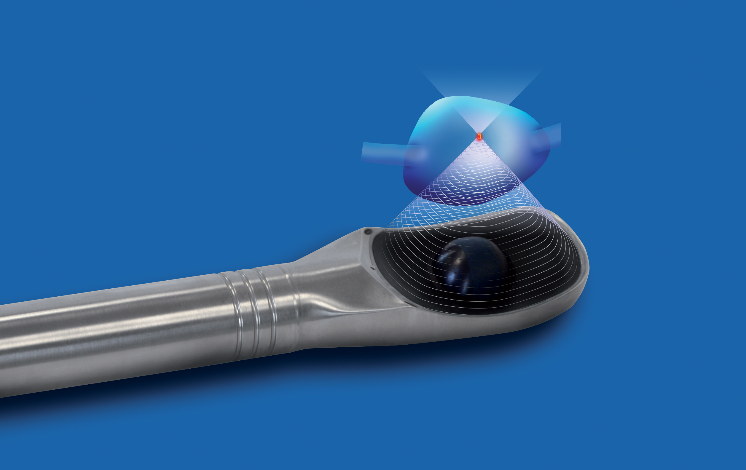
Radical prostatectomy and whole-gland radiation are reliable at controlling prostate cancer, but both carry a burden of side effects, including erectile and urinary dysfunction. Focal therapy emerged directly in response to this problem, offering an alternative for men with smaller, unifocal disease, aiming to ablate only the known tumor while sparing surrounding tissue and function.
The obstacle has rarely been the ablation technology itself. Adoption has been limited in part by how difficult it can be to precisely map significant cancer on MRI and identify appropriate patients. Tools like MSKCC-style calculators were built to flag which patients have significant disease at a population level, not to map a single lesion's exact borders in one gland. AI-enhanced nomograms are now being built to zero in on lesion location, tumor extent within the gland and the certainty behind that assessment.
Focal therapy requires a precise understanding of where the cancer is located within the prostate. AI imaging may provide additional information to help physicians determine whether a patient is an appropriate candidate.
-Dr. Julio Pow-Sang
How Accurately Can AI Identify and Map Prostate Cancer?
In one international study comparing AI detection systems against 62 radiologists reading the same MRI scans, the AI system detected 6.8% more clinically significant cancers while cutting false positives roughly in half. A separate deep learning model tested against four experienced radiologists produced a comparably higher accuracy score, with a false positive rate of 44% versus 71% for the radiologists, while catching cancer at nearly the same rate.
Mapping accuracy is just as critical as detection. One retrospective study used an FDA-cleared AI platform to generate three-dimensional cancer maps for 147 patients, then compared its extracapsular extension predictions against several established tools, including MRI scoring, Partin tables and the PReCEd nomogram, confirming results against surgical pathology. The AI mapping software outperformed every conventional method tested.
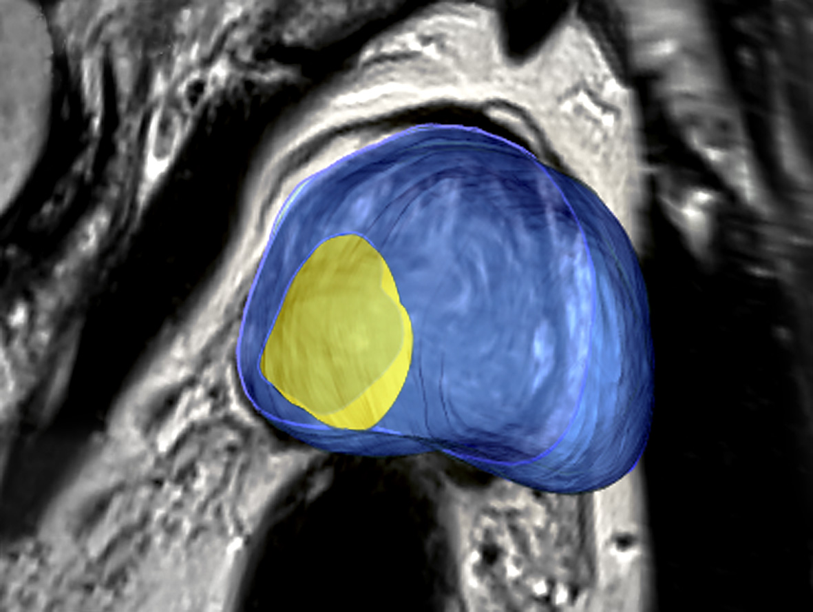 For focal therapy planning selection, this translates to:
- Greater confidence identifying unifocal, clinically significant disease
- Sharper lesion borders for more precise ablation margins
- Better pre-treatment risk stratification for extracapsular extension
What is Moffitt Contributing to This Research?
Moffitt's Department of Machine Learning has an active research program in prostate imaging, led in part by Yoga Balagurunathan, PhD in collaboration with genitourinary oncology chair Julio Pow-Sang, MD. That work includes a deep learning architecture that automatically segments the prostate gland and peripheral zone on MRI, along with ongoing research into the challenges of applying these models consistently across multiple readers. Both are foundational steps in the imaging-and-risk-stratification pipeline that any AI-assisted nomogram depends on.
Dr. Pow-Sang has spoken publicly about where the field is headed, noting that improvements in imaging, biopsy protocols, patient selection and follow-up will likely increase focal therapy use, while acknowledging that with five or six ablation technologies now available, it remains an open question which will ultimately prevail. He warns about the technology's current limits, too.
It is a promising technology, but it hasn't been proven long term. So, men have to be aware that they must be followed up closely."
-Dr. Julio Pow-Sang
Moffitt’s pathology and machine learning leadership, Marilyn Bui, MD, PhD, has described an institution-wide push toward full digital pathology adoption by 2027, with internal demand for AI algorithms and clinical utility in prostate and breast cancer, to feed AI-based risk models going forward.
What Referring Practitioners Should Take From This
Clinical judgment remains essential, but AI-enhanced nomograms are improving the quality of the information that guides those decisions. For patients with small, MRI-visible, unifocal tumors, these tools are helping clinicians more confidently identify candidates for focal therapy while better assessing the likelihood of extracapsular extension before treatment.
As the technology continues to evolve, multidisciplinary evaluation remains important. Questions about long-term outcomes, performance across diverse patient populations and optimal treatment selection are still being studied, making early collaboration with a specialized prostate cancer team valuable.
Referring patients to Moffitt before definitive treatment decisions are made allows for a comprehensive evaluation of all appropriate options. Through advanced MRI protocols, AI-assisted imaging analysis, multidisciplinary review and access to multiple focal therapy technologies alongside surgery, radiation and systemic therapies, Moffitt partners with referring physicians to determine the treatment approach that best aligns with each patient's disease and goals.
Have a patient who may be a candidate for focal therapy? To discuss treatment options for a patient with localized prostate cancer, complete the online referral form or email Physician.Relations@Moffitt.org.

 Medically reviewed by Julio Pow-Sang, MD, Department of Genitourinary Oncology.